
Page 629 - Small Animal Internal Medicine, 6th Edition
P. 629
CHAPTER 36 Hepatobiliary Diseases in the Dog 601
motility may also predispose to mucocele formation. A dyslipidemia in all cases, whether surgically or medically
mutation in biliary phosphatidylcholine transporter was managed.
VetBooks.ir proposed as a cause in affected Shetland Sheepdogs and also EXTRAHEPATIC BILE DUCT
a few dogs of other breeds with mucocele (Mealey et al.,
OBSTRUCTION
2010) but later disputed (Cullen et al., 2014).
Clinical signs vary. In some dogs mucocele is clinically The causes of EBDO in dogs are similar to those in cats (see
silent and is an incidental finding on abdominal ultrasonog- Box 35.4) with the exception of liver flukes, which are
raphy (see Fig. 36.8). In others nonspecific clinical signs are uncommon in dogs. The most common cause of EBDO in
seen, similar to those of other biliary tract diseases with dogs is extraluminal obstruction from acute-on-chronic
anorexia, lethargy, vomiting, and icterus. Some dogs present pancreatitis (see Chapter 37), but intestinal foreign bodies,
acutely because of gallbladder rupture and bile peritonitis. A neoplasia, bile duct involvement in a diaphragmatic hernia,
recent study demonstrated that ultrasonography has a high and other processes can also cause EBDO (Fig. 36.9). Bile
specificity but low sensitivity of only 56% for gallbladder duct injuries that heal and result in stricture formation
rupture and bile peritonitis for dogs with mucocele (Jaffey several weeks later are also seen in dogs; the common bile
et al., 2018). Therefore, if gallbladder rupture is suspected duct (CBD) may be compressed when carried with the liver
clinically, surgery should be undertaken even if the ultra- into the thorax in dogs with diaphragmatic hernia. Extralu-
sound is negative. minal compressive lesions, such as pancreatic, biliary, or
Treatment is usually surgical for clinically affected dogs; duodenal neoplasms, are less common causes, and choleli-
cholecystectomy with or without biliary diversion is the thiasis as a cause of EBDO is rare. To be considered as EBDO,
technique of choice. Biliary diversion increases perioperative a pathologic process must exist at the level of the CBD that
mortality and is rarely necessary. Gallbladder rupture also impedes bile flow into the duodenum. Only if bile flow has
increases the risk of death. However, those that survive the been completely interrupted for several weeks are acholic
perioperative period have a good long-term prognosis. feces, vitamin K–responsive coagulopathy, and repeated
Medical management of subclinical mucoceles has been absence of urobilinogen in properly processed urine speci-
reported in two dogs with hypothyroidism that were success- mens found. If obstruction is incomplete, these features are
fully treated (Walter et al., 2008) and in Shetland Sheepdogs not present and the constellation of signs and clinicopatho-
(Aguirre et al., 2007). In the shelties, medical management logic test results resembles those of other, nonobstructive
consisted of a low-fat diet (e.g., Hill’s i/d low fat; Royal Canin biliary tract disorders.
Waltham Gastrointestinal Low Fat; Eukanuba Intestinal
Diet, Procter & Gamble Pet Care, Mason, OH) with a cho- Clinical Features
leretic (ursodeoxycholic acid, 10–15 mg/kg PO total daily Presenting clinical signs and clinicopathologic and physical
dosage, preferably split twice) and antioxidant (SAM-e, examination findings of all these disorders may not differ
20 mg/kg PO q24h). In one dog this resulted in resolution greatly unless the underlying condition has caused EBDO
of the mucocele; in two dogs the mucocele remained static; or bile peritonitis. Regardless of the underlying disorder,
one dog died as a result of gallbladder rupture and one as a typical clinical signs are jaundice, acute or chronic vomiting,
result of pulmonary thromboembolism, both within 2 weeks anorexia, depression, weight loss, and occasionally vague
of diagnosis; and two dogs were lost to follow-up. It would cranial abdominal pain. Because of the protected location
also seem sensible to address the underlying cause of the of the gallbladder in the abdomen, it is rarely possible to be
A B
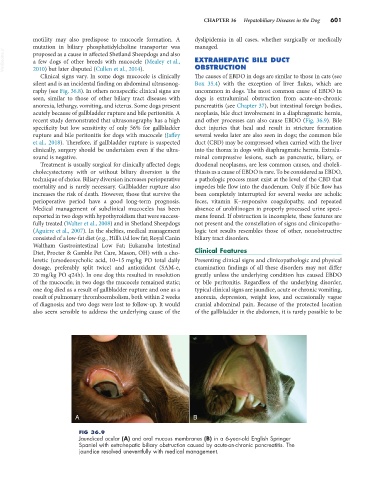
FIG 36.9
Jaundiced ocular (A) and oral mucous membranes (B) in a 6-year-old English Springer
Spaniel with extrahepatic biliary obstruction caused by acute-on-chronic pancreatitis. The
jaundice resolved uneventfully with medical management.