
Page 1280 - Veterinary Immunology, 10th Edition
P. 1280
and degeneration. Synovial biopsies show a neutrophil and
VetBooks.ir mononuclear cell infiltration with a fibrinous exudate. IgG, IgM,
and complement are deposited in the walls of the synovial vessels.
Animals may be treated with corticosteroids and
immunosuppressive agents such as cyclophosphamide.
Idiopathic Polyarthritis
Most cases of canine polyarthritis fit none of the categories
described previously. Although these cases are nonerosive and
possess the characteristics of type III hypersensitivity, their precise
etiology is unknown. They can be classified into four types (Table
38.1). Type I disease is polyarthritis alone. Type II disease is a
reactive arthritis associated with infections in the respiratory or
urinary tract, tooth infections, or cellulitis. Type III disease is
associated with the presence of gastroenteritis, diarrhea, or
ulcerative colitis. It is not clear whether this type of disease is truly
distinguishable from type II disease. Type IV disease is associated
with the presence of tumors, including seminomas and carcinomas.
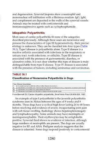
TABLE 38.1
Classification of Nonerosive Polyarthritis in Dogs
Type Disease Associations
I Uncomplicated polyarthritis without other disease associations
II Polyarthritis associated with infectious lesions remote from the joints (e.g., respiratory or urinary infections)
III Polyarthritis associated with gastrointestinal disease
IV Polyarthritis associated with neoplastic disease remote from the joints
From Bennett DJ: Canine idiopathic polyarthritis, Small Anim Pract 28:909-928, 1987.
An example of type I polyarthritis is the juvenile polyarthritis
syndrome seen in Akitas between the ages of 9 weeks and 8
months. These dogs have a cyclical high fever lasting 24 to 48 hours
before resolving and evidence of severe, incapacitating joint pain
with soft tissue swelling. Radiology shows hepatosplenomegaly
and lymphadenopathy. Some animals may have meningitis or
meningoencephalitis. Their erythrocytes may be antiglobulin
positive. Synovial fluid shows no evidence of infection, although
large numbers of neutrophils are present. The dogs are usually
negative for RF and ANA. Pedigree analysis suggests that the
disease is inherited. Some dogs respond positively to corticosteroid
1280