Page 1284 - Veterinary Immunology, 10th Edition
P. 1284
VetBooks.ir


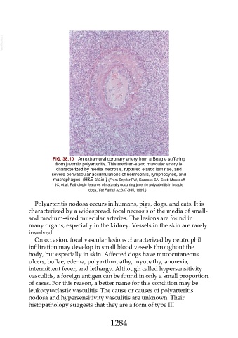
                           FIG. 38.10  An extramural coronary artery from a Beagle suffering
                             from juvenile polyarteritis. This medium-sized muscular artery is
                             characterized by medial necrosis, ruptured elastic laminae, and
                           severe perivascular accumulations of neutrophils, lymphocytes, and
                            macrophages. (H&E stain.) (From Snyder PW, Kazacos EA, Scott-Moncrieff
                            JC, et al: Pathologic features of naturally occurring juvenile polyarteritis in beagle
                                             dogs, Vet Pathol 32:337-345, 1995.)


                  Polyarteritis nodosa occurs in humans, pigs, dogs, and cats. It is
               characterized by a widespread, focal necrosis of the media of small-
               and medium-sized muscular arteries. The lesions are found in
               many organs, especially in the kidney. Vessels in the skin are rarely

               involved.
                  On occasion, focal vascular lesions characterized by neutrophil
               infiltration may develop in small blood vessels throughout the
               body, but especially in skin. Affected dogs have mucocutaneous

               ulcers, bullae, edema, polyarthropathy, myopathy, anorexia,
               intermittent fever, and lethargy. Although called hypersensitivity
               vasculitis, a foreign antigen can be found in only a small proportion
               of cases. For this reason, a better name for this condition may be

               leukocytoclastic vasculitis. The cause or causes of polyarteritis
               nodosa and hypersensitivity vasculitis are unknown. Their
               histopathology suggests that they are a form of type III


                                                        1284

