
Page 326 - The Veterinary Laboratory and Field Manual 3rd Edition
P. 326
Haematology 295
(ribs, pelvis and skull) and the short bones of the standard values and value ranges for each veteri-
vertebrae contain red marrow throughout life. nary laboratory. It is standard practice to list the
Bone marrow samples collected from freshly normal range of values alongside the test values
dead animals can provide useful and relatively to allow the submitting veterinary or livestock
easily collected diagnostic material but only extension officer to assess the results against
appropriately qualified and experienced veteri- the normal range for the laboratory. The normal
nary clinicians should collect bone marrow from
a live animal.
The most popular puncture site chosen in
live animals is the iliac crest or the head of the
femur (dog, cat) but the ribs and sternum may
also be used (cow, horse). Once the animal is
appropriately restrained (or under anaesthesia),
local anaesthetic (for example, lignocaine) is
injected around the region and a skin incision
made over the selected bone. A sterile wide bore
needle is required to aspirate the marrow mate-
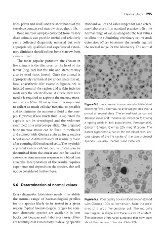
rial using a 10 or 20 ml syringe. It is important Figure 5.6 Blood smear from a cow which later died
to collect as much cellular material as possible following fever, haematuria and weight loss over a
and to minimize the amount of fluid in the sam- period of several days. The animal had concurrent
ple. However, if too much fluid is aspirated the Babesia bovis and Theileria sp. infection following
aspirate can be centrifuged and the sediment a spring peak in tick populations, Pemagatshel,
examined on a microscope slide. The prepared Eastern Bhutan. Giemsa 20x magnification. The
bone marrow smear can be fixed in methanol darker pigmented areas in the red blood cells indi-
and stained with Giemsa stain as for a routine cate stages of the life cycles of the two protozoal
blood smear. A differential count is usually made species. See also Chapter 3 and Plate 22a.
after counting 500 nucleated cells. The myeloid/
erythroid (white cell/red cell) ratio can also be
determined from the smear and can be used to
assess the bone marrow response to a blood loss
anaemia. Interpretation of the results requires
experience and depends on the species, this will
not be considered further here.
5.6 determination of normal values
Every diagnostic laboratory needs to establish
the normal range of haematological profiles Figure 5.7 Poor quality bovine blood smear stained
for the species likely to be tested in a given with Giemsa 100× oil immersion. Note the pres-
region. Typical haematological ranges for com- ence of a large mononuclear cell. The red cells
mon domestic species are available in text are irregular in shape and there is a lot of artefact.
books but because each laboratory uses differ- The presence of granules suggests that new stain
ent techniques it is necessary to develop specific should be prepared. See also Plate 22b.
Vet Lab.indb 295 26/03/2019 10:25