
Page 328 - The Veterinary Laboratory and Field Manual 3rd Edition
P. 328
Haematology 297
(a) (c)
(b)
(d)
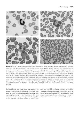
Figure 5.10 (a) Canine band neutrophil (immature PMN). Note the band shaped nucleus with smooth
sides and lack of constrictions. This cell also shows signs of toxic change with a foamy appearance due
to the presence of vacuoles. Modified Wright’s stain 100×. (b) Feline eosinophil. Note reddish granules in
the cytoplasm and segmented nucleus. The nuclear segments are connected by a fine strand. Wright’s
stain 100×. (c) Feline basophil. Note pale lavender granules in the cytoplasm and segmented nucleus. The
colour of the cytoplasmic granules varies from pale grey to dark purple depending on the species. Wright’s
stain 100×. (d) Feline blood smear showing rouleaux formation. This is commonly seen in equine and
feline blood smears. Wright’s stain 20×. See also Plates 25, 26, 27 and 28. Photos: Dr Nicole Fernandez,
WCVM.
ist knowledge and experience are required to are any suitable training courses available.
assess more subtle changes in the blood pic- Additional information can be found in the texts
ture. In order to learn more about the topic it is listed in the bibliography and on websites, such
advisable to speak to the laboratory manager as the Cornell University Hematology Atlas.
5
or the regional veterinary officer to see if there
Vet Lab.indb 297 26/03/2019 10:26