
Page 352 - The Veterinary Laboratory and Field Manual 3rd Edition
P. 352
Serology and immunology 321
cein will take place and this can be visualized
under a fluorescent microscope. This test is more
sensitive than the direct test and fluorescence is
often brighter because there are more combining
sites for the fluorescent anti-immunoglobulin.
These tests should be carried out in accor-
dance with instructions provided with the
reagents. To ensure that the test performs well
the following should be considered.
• Make thin smears of material under test.
• Stick rigidly to pH and incubation recommen-
dations.
• Do not use the fluorescence microscope if the
voltage is fluctuating.
• Do not use too much mounting fluid and
watch for air bubbles.
• Run positive and negative controls with each
test.
• Do not allow preparations to dry out.
The test can also be performed using non-fluo-
rescent labels, such as horseradish peroxidase
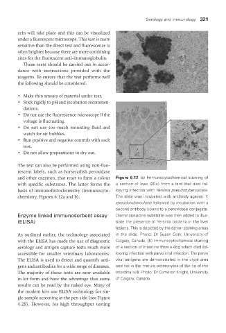
and other enzymes, that react to form a colour Figure 6.12 (a) Immunocytochemical staining of
with specific substrates. The latter forms the a section of liver (20×) from a bird that died fol-
basis of immunohistochemistry (immunocyto- lowing infection with Yersinia pseudotuberculosis.
chemistry, Figures 6.12a and b). The slide was incubated with antibody against Y.
pseudotuberculosis followed by incubation with a
second antibody bound to a peroxidase conjugate.
Enzyme linked immunosorbent assay Diaminobezidine substrate was then added to illus-
(ELISa) trate the presence of Yersinia bacteria in the liver
lesions. This is depicted by the darker staining areas
As outlined earlier, the technology associated in the slide. Photo: Dr Susan Cork, University of
with the ELISA has made the use of diagnostic Calgary, Canada. (b) Immunocytochemical staining
serology and antigen capture tests much more of a section of intestine from a dog which died fol-
accessible for smaller veterinary laboratories. lowing infection with parvo viral infection. The parvo
The ELISA is used to detect and quantify anti- viral antigens are demonstrated in the crypt area
gens and antibodies for a wide range of diseases. and not in the mature enterocytes of the tip of the
The majority of these tests are now available intestinal villi. Photo: Dr Cameron Knight, University
in kit form and have the advantage that some of Calgary, Canada.
results can be read by the naked eye. Many of
the modern kits use ELISA technology for sin-
gle sample screening at the pen side (see Figure
4.29). However, for high throughput testing
Vet Lab.indb 321 26/03/2019 10:26