
Page 882 - Problem-Based Feline Medicine
P. 882
874 PART 10 CAT WITH SIGNS OF NEUROLOGICAL DISEASE
A
D
C
B
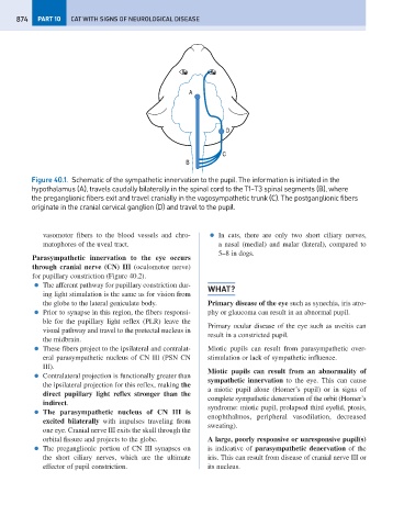
Figure 40.1. Schematic of the sympathetic innervation to the pupil. The information is initiated in the
hypothalamus (A), travels caudally bilaterally in the spinal cord to the T1–T3 spinal segments (B), where
the preganglionic fibers exit and travel cranially in the vagosympathetic trunk (C). The postganglionic fibers
originate in the cranial cervical ganglion (D) and travel to the pupil.
vasomotor fibers to the blood vessels and chro- ● In cats, there are only two short ciliary nerves,
matophores of the uveal tract. a nasal (medial) and malar (lateral), compared to
5–8 in dogs.
Parasympathetic innervation to the eye occurs
through cranial nerve (CN) III (oculomotor nerve)
for pupillary constriction (Figure 40.2).
● The afferent pathway for pupillary constriction dur-
WHAT?
ing light stimulation is the same as for vision from
the globe to the lateral geniculate body. Primary disease of the eye such as synechia, iris atro-
● Prior to synapse in this region, the fibers responsi- phy or glaucoma can result in an abnormal pupil.
ble for the pupillary light reflex (PLR) leave the
Primary ocular disease of the eye such as uveitis can
visual pathway and travel to the pretectal nucleus in
result in a constricted pupil.
the midbrain.
● These fibers project to the ipsilateral and contralat- Miotic pupils can result from parasympathetic over-
eral parasympathetic nucleus of CN III (PSN CN stimulation or lack of sympathetic influence.
III).
Miotic pupils can result from an abnormality of
● Contralateral projection is functionally greater than
sympathetic innervation to the eye. This can cause
the ipsilateral projection for this reflex, making the
a miotic pupil alone (Horner’s pupil) or in signs of
direct pupillary light reflex stronger than the
complete sympathetic denervation of the orbit (Horner’s
indirect.
syndrome: miotic pupil, prolapsed third eyelid, ptosis,
● The parasympathetic nucleus of CN III is
enophthalmos, peripheral vasodilation, decreased
excited bilaterally with impulses traveling from
sweating).
one eye. Cranial nerve III exits the skull through the
orbital fissure and projects to the globe. A large, poorly responsive or unresponsive pupil(s)
● The preganglionic portion of CN III synapses on is indicative of parasympathetic denervation of the
the short ciliary nerves, which are the ultimate iris. This can result from disease of cranial nerve III or
effector of pupil constriction. its nucleus.