
Page 883 - Problem-Based Feline Medicine
P. 883
40 – THE CAT WITH ANISOCORIA OR ABNORMALLY DILATED OR CONSTRICTED PUPILS 875
A
B
D D
C
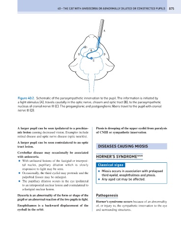
Figure 40.2. Schematic of the parasympathetic innervation to the pupil. The information is initiated by
a light stimulus (A), travels caudally in the optic nerve, chiasm and optic tract (B), to the parasympathetic
nucleus of cranial nerve III (C). The preganglionic and postganglionic fibers travel to the pupil with cranial
nerve III (D).
A larger pupil can be seen ipsilateral to a prechias- Ptosis is drooping of the upper eyelid from paralysis
mic lesion causing decreased vision. Examples include of CNIII or sympathetic innervation.
retinal disease and optic nerve disease (optic neuritis).
A larger pupil can be seen contralateral to an optic
tract lesion. DISEASES CAUSING MIOSIS
Cerebellar disease may occasionally be associated
with anisocoria. HORNER’S SYNDROME***
● With unilateral lesions of the fastigial or interposi-
tal nuclei, pupillary dilation which is slowly Classical signs
responsive to light may be seen.
● Miosis occurs in association with prolapsed
● Occasionally, the third eyelid may protrude and the
third eyelid, enophthalmos and ptosis.
palpebral fissure may be enlarged.
● Any aged cat may be affected.
● The pupillary dilation occurs in the eye ipsilateral
to an interposital nuclear lesion and contralateral to
a fastigial nuclear lesion.
Dyscoria is an abnormality of the form or shape of the Pathogenesis
pupil or an abnormal reaction of the two pupils to light.
Horner’s syndrome occurs because of an abnormality
Enophthamos is a backward displacement of the of, or injury to, the sympathetic innervation to the eye
eyeball in the orbit. and surrounding structures.