
Page 1010 - Adams and Stashak's Lameness in Horses, 7th Edition
P. 1010
976 Chapter 9
Intra‐articular chip fractures of the dorsal aspect of relatively common in the Quarter horse, despite such
the proximal phalanx are commonly seen in the fore fractures being considered fatigue‐related injuries by
VetBooks.ir fragments). These fractures are considered to be trau Thoroughbred racing, biaxial and comminuted sesa
other authors. Although most often associated with
limb of racing Quarter horses (but less than carpal chip
matic hyperextension injuries. They occur primarily on
moid fractures occur in the Quarter horse, resulting in
8
the medial aspect, but may also occur laterally. In some disruption of the suspensory apparatus.
cases, they are quite large, especially compared to the
fragments that occur in the Thoroughbred, with a long Dorsal Metacarpal Disease
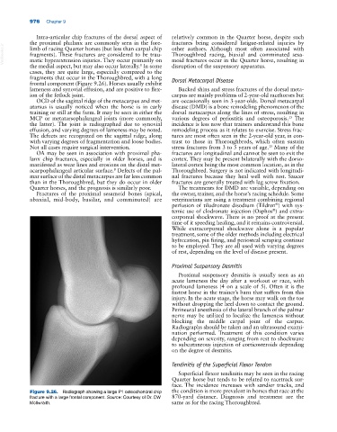
frontal component (Figure 9.26). Horses usually exhibit
lameness and synovial effusion, and are positive to flex Bucked shins and stress fractures of the dorsal meta
ion of the fetlock joint. carpus are mainly problems of 2‐year‐old racehorses but
OCD of the sagittal ridge of the metacarpus and met are occasionally seen in 3‐year‐olds. Dorsal metacarpal
atarsus is usually noticed when the horse is in early disease (DMD) is a bone remodeling phenomenon of the
training or still at the farm. It may be seen in either the dorsal metacarpus along the lines of stress, resulting in
MCP or metatarsophalangeal joints (more commonly, various degrees of periostitis and osteoporosis. The
15
the latter). The joint is radiographed due to synovial incidence is less now that trainers understand this bone
effusion, and varying degrees of lameness may be noted. remodeling process as it relates to exercise. Stress frac
The defects are recognized on the sagittal ridge, along tures are most often seen in the 2‐year‐old year, in con
with varying degrees of fragmentation and loose bodies. trast to those in Thoroughbreds, which often sustain
15
Not all cases require surgical intervention. stress fractures from 3 to 5 years of age. Many of the
OA may be seen in association with proximal pha fractures are longitudinal and cannot be seen to exit the
lanx chip fractures, especially in older horses, and is cortex. They may be present bilaterally with the dorso
manifested as wear lines and erosions on the distal met lateral cortex being the most common location, as in the
acarpophalangeal articular surface. Defects of the pal Thoroughbred. Surgery is not indicated with longitudi
8
mar surface of the distal metacarpus are far less common nal fractures because they heal well with rest. Saucer
than in the Thoroughbred, but they do occur in older fractures are generally treated with lag screw fixation.
Quarter horses, and the prognosis is similarly poor. The treatments for DMD are variable, depending on
Fractures of the proximal sesamoid bones (apical, the owner, trainer, and the horse’s racing schedule. Some
abaxial, mid‐body, basilar, and comminuted) are veterinarians are using a treatment combining regional
perfusion of tiludronate disodium (Tildren ) with sys
®
temic use of clodronate injection (Osphos ) and extra
®
corporeal shockwave. There is no proof at the present
time of it speeding healing, and it remains controversial.
While extracorporeal shockwave alone is a popular
treatment, some of the older methods including electrical
hyfrecation, pin firing, and periosteal scraping continue
to be employed. They are all used with varying degrees
of rest, depending on the level of disease present.
Proximal Suspensory Desmitis
Proximal suspensory desmitis is usually seen as an
acute lameness the day after a workout or race, with
profound lameness (4 on a scale of 5). Often it is the
fastest horse in the trainer’s barn that suffers from this
injury. In the acute stage, the horse may walk on the toe
without dropping the heel down to contact the ground.
Perineural anesthesia of the lateral branch of the palmar
nerve may be utilized to localize the lameness without
blocking the middle carpal joint of the carpus.
Radiographs should be taken and an ultrasound exami
nation performed. Treatment of this condition varies
depending on severity, ranging from rest to shockwave
to subcutaneous injection of corticosteroids depending
on the degree of desmitis.
Tendinitis of the Superficial Flexor Tendon
Superficial flexor tendinitis may be seen in the racing
Quarter horse but tends to be related to racetrack sur
face. The incidence increases with sandier tracks, and
Figure 9.26. Radiograph showing a large P1 osteochondral chip the condition is more prevalent in horses that race at the
fracture with a large frontal component. Source: Courtesy of Dr. CW 870‐yard distance. Diagnosis and treatment are the
McIlwraith. same as for the racing Thoroughbred.