
Page 1011 - Adams and Stashak's Lameness in Horses, 7th Edition
P. 1011
Occupational‐Related Lameness Conditions 977
Arthrosis of the Carpus The diagnosis is generally made by physical examina
tion and digital radiography. The lameness is fairly
Carpal synovitis is the most frequent condition seen
VetBooks.ir in the young racing Quarter horse. Back‐in‐the‐knee ment or circumduction of the involved limb at the walk.
obvious; most horses have a characteristic wide place
conformation is common, and this predisposes to carpal
Most cases are sensitive to flexion of the carpus and pal
injury during hyperextension of the joint while running.
Many 2‐year‐olds have a large body mass for their age pation of the dorsal surface of the carpal bones. Heat
and synovial effusion are often present.
and reach very fast speeds without much prior condi Arthroscopic surgery is the treatment of choice.
tioning. The condition is characterized by heat and syn Distal radial carpal chips are particularly associated
ovial effusion of the affected joints with the absence of with progressive cartilage damage and OA if the horse
radiographic changes. Lameness may be present but is continues to race without removal of the chip fracture.
generally not severe. Carpal flexion and palpation are Many Quarter horses have multiple surgeries during
used to localize the affected joints. The treatment is the their racing careers due to the high incidence of osteo
same as for synovitis of the MCP joint.
chondral chip fragmentation.
Other important injuries include the more severe
fractures (slab fractures and comminuted fractures),
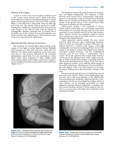
Osteochondral Chip Fractures of the Carpus many of which are amenable to surgery. Whenever sig
The incidence of osteochondral chip fractures of the nificant lameness is evident in the carpus, a thorough
carpus is very high in racing Quarter horses. Multiple radiographic examination including skyline views of the
chip fractures are often seen and many times they are carpus is necessary to ensure that degenerative lesions,
bilateral. The distal radial carpal bone is the most com incomplete sagittal fractures, and any nondisplaced
mon site for chip fracture, followed by the proximal fractures may be seen before a catastrophic injury
intermediate carpal bone. It is not uncommon to have occurs. All of the reported third carpal bone fractures
distal radial carpal fragments in both middle carpal can be seen in the Quarter horse, with a higher percent
joints and proximal intermediate carpal bone fragmen age of large frontal slab fractures involving both the
tation in both antebrachiocarpal joints in the same horse radial and intermediate facets (Figure 9.28). The slab is
(Figure 9.27). 13 often displaced in these cases, with the distal margin of
the radial carpal bone collapsing into the proximal frac
ture site (Figure 9.29). Lag screw fixation is necessary
for proper healing and to prevent further collapse of the
joint.
Fractures involving both rows of carpal bones may be
seen with some injuries. These can be challenging surgi
cal cases due to the severe damage to the carpal bones
and associated joint instability, requiring partial or pan‐
arthrodesis and the use of multiple bone plates. Most of
these injuries can be managed successfully by using
aggressive internal fixation techniques to produce pas
ture‐sound breeding animals. Prompt surgical interven
tion is necessary due to the high risk of laminitis in the
contralateral limb. 9
Figure 9.27. Radiograph demonstrating osteochondral chip
fragments off the proximal intermediate and distal radial carpal Figure 9.28. Skyline view of the third carpal bone with a slab
bones (a common combination in the racing Quarter horse). fracture involving both radial and intermediate facets. Source:
Source: Courtesy of Dr. CW McIlwraith. Courtesy of Dr. CW McIlwraith.