
Page 1012 - Adams and Stashak's Lameness in Horses, 7th Edition
P. 1012
978 Chapter 9
Hock Lameness If methylprednisolone acetate is used, low doses should
be considered (20–40 mg).
Effusion of the tarsocrural joint in the hock may be
VetBooks.ir indicative of OCD lesions associated primarily with Stifle Lameness
the distal intermediate ridge or medial malleolus of
the tibia. Often these cases are operated before arriv
ing at the track. Lameness associated with the distal The most common site of stifle pain is the medial
tarsal joints, as well as other hind end lameness, is femorotibial joint, which is the same in racing
associated with a failure to break sharply from the Thoroughbreds. This can be another cause of poor per
starting gate. Upper hindlimb flexion tests may be formance. The horse may be positive to upper hindlimb
equivocal, but a positive Churchill test along with the flexion or palpation, but most often intra‐articular anes
history of a poor performance may indicate tarsitis, thesia is required to localize the lameness. It is not
although a negative test does not rule out the prob always clear whether the condition is synovitis or early
lem. The condition is generally bilateral, and the horse OA. Radiographs are useful to assess the joint and rule
may track close behind or cross midline when out certain conditions. Ultrasound examination is useful
observed from behind. to pick up soft tissue conditions. 4
Horses that wear patches behind to protect from Most stifle lameness responds well to intra‐articular
scalping or horses with laceration marks seen on the therapy; however, if lameness persists, diagnostic arthros
medial aspect of the hock are highly suspect for hock copy is necessary to make a definitive diagnosis. Lameness
soreness. Radiographs are negative in many instances, conditions involving the femoropatellar joint are often
or subtle changes may be seen. Intra‐articular corticos accompanied by the presence of joint effusion, but in recent
teroids are effective at relieving the lameness and/or years most OCD lesions are operated on before the horse
improving performance. Even though the tarsometatar starts racing. Upward fixation of the patella can be an issue
sal and centrodistal joints are low‐motion joints, main in immature racehorses in early training. Subchondral
taining articular cartilage is important because these cystic lesions of the medial femoral condyle are a relatively
joints rarely fuse on their own. Betamethasone esters rare but painful condition in the racehorse.
(Betavet ) and triamcinolone acetate (Vetalog ) have
®
®
been shown to have fewer deleterious effects on carti Tibial Stress Fractures
lage than methylprednisolone acetate (Depo‐Medrol ).
®
5,6
Stress fractures involving the tibia are seen in young
horses around the time of their second qualifying work
or first race. Diagnosis of the condition has become
more common with the advances in imaging techniques
and the access to nuclear scintigraphy. The lameness is
unilateral and quite severe; the left hindleg is predominantly
affected. This may be due to the fact that the horse pulls
up quickly from high speed before entering a left‐hand
turn on the racetrack. If a tibial stress fracture is sus
pected, nuclear scintigraphy is the best way to demon
strate the injury. Alternatively, digital radiographs taken
1 week to 10 days after injury may show a lesion on the
tibia. The horse is usually rested for at least 90 days
before resuming training.
Catastrophic Fractures
Proximal sesamoid fractures are the greatest cause of
catastrophic injury in the racing Quarter horse. A retro
spective study of racing fatalities found that carpal bone
and vertebral body fractures were more common in
Quarter horses than Thoroughbred racehorses. Sprinting
vs. distance racing may play a role in a different distri
bution of skeletal injuries, but the greatest cause of death
for both breeds was found to be fetlock injury. 16
Lumbar vertebral fractures of the spine have been
associated with serious injury to jockeys due to the fall
of horse and rider. 3,11 The fractures are located at the
L5–L6 vertebral junction. Pathology of the lumbar
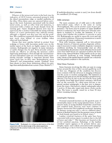
Figure 9.29. Radiograph of a collapsing slab fracture of the third vertebral region appears to be present before cata
carpal bone with the distal margin of the radial carpal bone strophic injury and is likely associated with maladap
collapsing into the proximal fracture site. Source: Courtesy of Dr. tive pathology at the L5–L6 location (Figure 9.30). 3
CW McIlwraith.