
Page 303 - Adams and Stashak's Lameness in Horses, 7th Edition
P. 303
VetBooks.ir
1
2
a
c 3
4
27 5
26
25 6
b
24
23 7
22
8
21
20 d 9
19 10
18 e 11
12
17
13
16 f 14
15
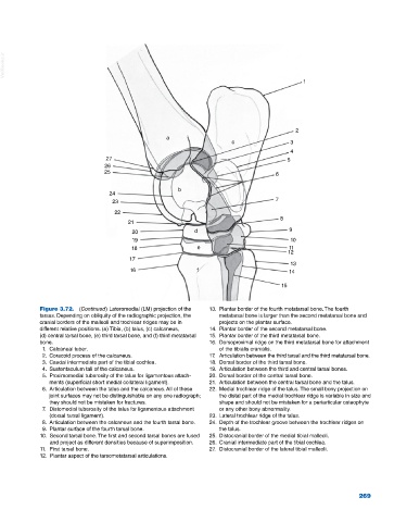
Figure 3.72. (Continued) Lateromedial (LM) projection of the 13. Plantar border of the fourth metatarsal bone. The fourth
tarsus. Depending on obliquity of the radiographic projection, the metatarsal bone is larger than the second metatarsal bone and
cranial borders of the malleoli and trochlear ridges may be in projects on the plantar surface.
different relative positions. (a) Tibia, (b) talus, (c) calcaneus, 14. Plantar border of the second metatarsal bone.
(d) central tarsal bone, (e) third tarsal bone, and (f) third metatarsal 15. Plantar border of the third metatarsal bone.
bone. 16. Dorsoproximal ridge on the third metatarsal bone for attachment
1. Calcaneal tuber. of the tibialis cranialis.
2. Coracoid process of the calcaneus. 17. Articulation between the third tarsal and the third metatarsal bone.
3. Caudal intermediate part of the tibial cochlea. 18. Dorsal border of the third tarsal bone.
4. Sustentaculum tali of the calcaneus. 19. Articulation between the third and central tarsal bones.
5. Proximomedial tuberosity of the talus for ligamentous attach 20. Dorsal border of the central tarsal bone.
ments (superficial short medial collateral ligament). 21. Articulation between the central tarsal bone and the talus.
6. Articulation between the talus and the calcaneus. All of these 22. Medial trochlear ridge of the talus. The small bony projection on
joint surfaces may not be distinguishable on any one radiograph; the distal part of the medial trochlear ridge is variable in size and
they should not be mistaken for fractures. shape and should not be mistaken for a periarticular osteophyte
7. Distomedial tuberosity of the talus for ligamentous attachment or any other bony abnormality.
(dorsal tarsal ligament). 23. Lateral trochlear ridge of the talus.
8. Articulation between the calcaneus and the fourth tarsal bone. 24. Depth of the trochlear groove between the trochlear ridges on
9. Plantar surface of the fourth tarsal bone. the talus.
10. Second tarsal bone. The first and second tarsal bones are fused 25. Distocranial border of the medial tibial malleoli.
and project as different densities because of superimposition. 26. Cranial intermediate part of the tibial cochlea.
11. First tarsal bone. 27. Distocranial border of the lateral tibial malleoli.
12. Plantar aspect of the tarsometatarsal articulations.
269