
Page 305 - Adams and Stashak's Lameness in Horses, 7th Edition
P. 305
VetBooks.ir 1
2
a 3
4
24 5
23 6
b
e d
7
f
c
8
9
13 12 11 10
22 21 20 19 18 17 16 15 14
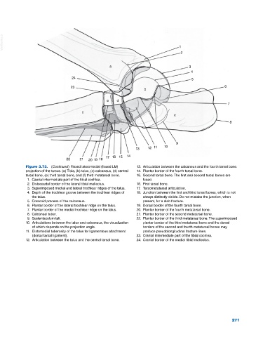
Figure 3.73. (Continued) Flexed lateromedial (flexed LM) 13. Articulation between the calcaneus and the fourth tarsal bone.
projection of the tarsus. (a) Tibia, (b) talus, (c) calcaneus, (d) central 14. Plantar border of the fourth tarsal bone.
tarsal bone, (e) third tarsal bone, and (f) third metatarsal bone. 15. Second tarsal bone. The first and second tarsal bones are
1. Caudal intermediate part of the tibial cochlea. fused.
2. Distocaudal border of the lateral tibial malleolus. 16. First tarsal bone.
3. Superimposed medial and lateral trochlear ridges of the talus. 17. Tarsometatarsal articulation.
4. Depth of the trochlear groove between the trochlear ridges of 18. Junction between the first and third tarsal bones, which is not
the talus. always distinctly visible. Do not mistake the junction, when
5. Coracoid process of the calcaneus. present, for a slab fracture.
6. Plantar border of the lateral trochlear ridge on the talus. 19. Dorsal border of the fourth tarsal bone.
7. Plantar border of the medial trochlear ridge on the talus. 20. Plantar border of the fourth metatarsal bone.
8. Calcaneal tuber. 21. Plantar border of the second metatarsal bone.
9. Sustentaculum tali. 22. Plantar border of the third metatarsal bone. The superimposed
10. Articulations between the talus and calcaneus, the visualization plantar border of the third metatarsal bone and the dorsal
of which depends on the projection angle. borders of the second and fourth metatarsal bones may
11. Distomedial tuberosity of the talus for ligamentous attachment produce pseudolongitudinal fracture lines.
(dorsal tarsal ligament). 23. Cranial intermediate part of the tibial cochlea.
12. Articulation between the talus and the central tarsal bone. 24. Cranial border of the medial tibial malleolus.
271