
Page 307 - Adams and Stashak's Lameness in Horses, 7th Edition
P. 307
VetBooks.ir 32
a
31
1
30 b
2
29 3
4
28
5
27
6
c 7
26 8
25 9
24
23 10
22 11
11 12
12 13
14 14
21 15
20 d 16
19 17
e f
16
18
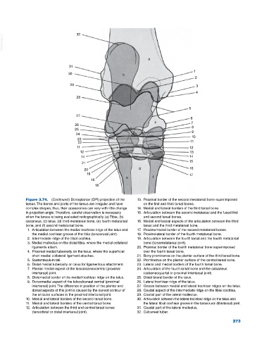
Figure 3.74. (Continued) Dorsoplantar (DPl) projection of the 13. Proximal border of the second metatarsal bone superimposed
tarsus. The bones and joints of the tarsus are irregular and have on the first and third tarsal bones.
complex shapes; thus, their appearance can vary with little change 14. Medial and lateral borders of the third tarsal bone.
in projection angle. Therefore, careful observation is necessary 15. Articulation between the second metatarsal and the fused first
when the tarsus is being evaluated radiographically. (a) Tibia, (b) and second tarsal bones.
calcaneus, (c) talus, (d) third metatarsal bone, (e) fourth metatarsal 16. Medial and lateral aspects of the articulation between the third
bone, and (f) second metatarsal bone. tarsal and the third metatarsal bone.
1. Articulation between the medial trochlear ridge of the talus and 17. Proximomedial border of the second metatarsal bones.
the medial cochlear groove of the tibia (tarsocrural joint). 18. Proximolateral border of the fourth metatarsal bone.
2. Intermediate ridge of the tibial cochlea. 19. Articulation between the fourth tarsal and the fourth metatarsal
3. Medial malleolus on the distal tibia, where the medial collateral bone (tarsometatarsal joint).
ligaments attach. 20. Proximal border of the fourth metatarsal bone superimposed
4. Proximal medial tuberosity on the talus, where the superficial over the fourth tarsal bone.
short medial collateral ligament attaches. 21. Bony prominence on the plantar surface of the third tarsal bone.
5. Sustentaculum tali. 22. Prominence on the plantar surface of the central tarsal bone.
6. Distal medial tuberosity on talus for ligamentous attachment. 23. Lateral and medial borders of the fourth tarsal bone.
7. Plantar medial aspect of the talocalcaneocentral (proximal 24. Articulation of the fourth tarsal bone and the calcaneus
intertarsal) joint. (calceneoquartal or proximal intertarsal joint).
8. Distomedial border of the medial trochlear ridge on the talus. 25. Distal lateral border of the talus.
9. Dorsomedial aspect of the talocalcaneal central (proximal 26. Lateral trochlear ridge of the talus.
intertarsal) joint. The difference in position of the plantar and 27. Groove between medial and lateral trochlear ridges on the talus.
dorsal aspects of this joint is caused by the curved contour of 28. Caudal aspect of the intermediate ridge on the tibial cochlea.
the articular surfaces in the proximal intertarsal joint. 29. Cranial part of the lateral malleolus.
10. Medial and lateral borders of the second tarsal bone. 30. Articulation between the lateral trochlear ridge on the talus and
11. Medial and lateral borders of the central tarsal bone. the lateral tibial cochlear groove in the tarsocrural (tibiotarsal) joint.
12. Articulation between the third and central tarsal bones 31. Caudal part of the lateral malleolus.
(tarsodistal or distal intertarsal joint). 32. Calcaneal tuber.
273