
Page 316 - Adams and Stashak's Lameness in Horses, 7th Edition
P. 316
VetBooks.ir
5
1
6
2 7
3 8
4
9
10
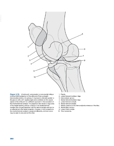
Figure 3.79. (Continued) Laterocaudal to craniomedial oblique 1. Patella.
(L45Ca‐CrMO) projection of the stifle joint. From a straight 2. Lateral femoral trochlear ridge.
lateromedial position, the primary X‐ray beam is directed parallel to 3. Intercondylar groove.
the ground with a 45° caudal angle and is centered on the caudal 4. Medial femoral trochlear ridge.
aspect of the stifle joint for a different approach in the evaluation of 5. Lateral femoral condyle.
the medial femoral condyle. This projection also shows a large area 6. Medial femoral condyle.
of the weight‐bearing articular surface of the medial femoral 7. Medial tubercle on the intercondyloid eminence of the tibia.
condyle free of superimposition with the lateral condyle and can be 8. Medial tibial condyle.
an alternative to the flexed projection. However, in both projections, 9. Lateral tibial condyle.
the medial femoral condyle is projected differently, and one lesion 10. Tibial tuberosity.
may be seen in one and not the other.
282