
Page 319 - Adams and Stashak's Lameness in Horses, 7th Edition
P. 319
33
VetBooks.ir 32 a 1
31
2
30
3
29 b 4
28
27
26 5
25
24 6
23
22 7
21 8
20
19 9
18 10
17
16
15
c
d
11
14
13
12
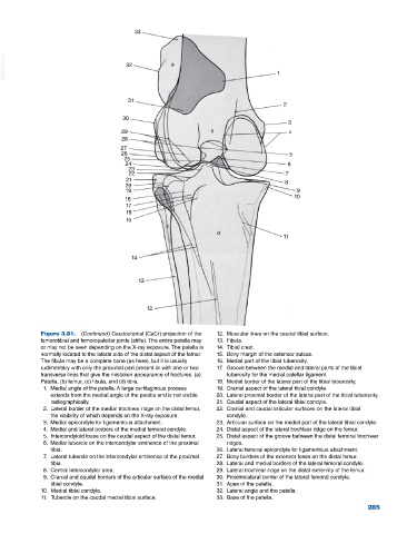
Figure 3.81. (Continued) Caudocranial (CaCr) projection of the 12. Muscular lines on the caudal tibial surface.
femorotibial and femoropatellar joints (stifle). The entire patella may 13. Fibula.
or may not be seen depending on the X‐ray exposure. The patella is 14. Tibial crest.
normally located to the lateral side of the distal aspect of the femur. 15. Bony margin of the extensor sulcus.
The fibula may be a complete bone (as here), but it is usually 16. Medial part of the tibial tuberosity.
rudimentary with only the proximal part present or with one or two 17. Groove between the medial and lateral parts of the tibial
transverse lines that give the mistaken appearance of fractures. (a) tuberosity for the medial patellar ligament.
Patella, (b) femur, (c) fibula, and (d) tibia. 18. Medial border of the lateral part of the tibial tuberosity.
1. Medial angle of the patella. A large cartilaginous process 19. Cranial aspect of the lateral tibial condyle.
extends from the medial angle of the patella and is not visible 20. Lateral proximal border of the lateral part of the tibial tuberosity.
radiographically. 21. Caudal aspect of the lateral tibial condyle.
2. Lateral border of the medial trochlear ridge on the distal femur, 22. Cranial and caudal articular surfaces on the lateral tibial
the visibility of which depends on the X‐ray exposure. condyle.
3. Medial epicondyle for ligamentous attachment. 23. Articular surface on the medial part of the lateral tibial condyle.
4. Medial and lateral borders of the medial femoral condyle. 24. Distal aspect of the lateral trochlear ridge on the femur.
5. Intercondyloid fossa on the caudal aspect of the distal femur. 25. Distal aspect of the groove between the distal femoral trochlear
6. Medial tubercle on the intercondylar eminence of the proximal ridges.
tibia. 26. Lateral femoral epicondyle for ligamentous attachment.
7. Lateral tubercle on the intercondylar eminence of the proximal 27. Bony borders of the extensor fossa on the distal femur.
tibia. 28. Lateral and medial borders of the lateral femoral condyle.
8. Central intercondylar area. 29. Lateral trochlear ridge on the distal extremity of the femur.
9. Cranial and caudal borders of the articular surface of the medial 30. Proximolateral border of the lateral femoral condyle.
tibial condyle. 31. Apex of the patella.
10. Medial tibial condyle. 32. Lateral angle and the patella.
11. Tubercle on the caudal medial tibial surface. 33. Base of the patella.
285