
Page 491 - Adams and Stashak's Lameness in Horses, 7th Edition
P. 491
Lameness of the Distal Limb 457
dorsal border lesions, sagittal plane and oblique splits, has not been documented. In many cases, however, it
and insertional lesions. 105,106 Abnormalities have been would seem likely that previous chronic abnormalities
VetBooks.ir (least common), the level of the navicular bone or CSLs addition, chronic DDFT injuries are not always isolated
of the DDFT would predispose to complete failure. In
identified at the DDFT insertion to the distal phalanx
to a single site (Figure 4.21), and there is evidence to
(most common), more proximally in the pastern, or a
combination of sites. 38,39,41–44,78 The most common loca- suggest that DDFT lesions may be a degenerative pro-
tion is at the level of the navicular bone and CSLs and cess due to vascular compromise rather than an inflam-
can be true core lesions, sagittal splits, or dorsal abra- matory process from trauma. 11
sions (Figure 4.13). 10,11,39,44 DDFT lesions can occur in a
single or multiple locations and can extend variable dis- Clinical Signs
tances up or down the tendon (Figure 4.21).
Abnormalities of the DDFT at the level of the proximal In general, horses that have primary soft tissue inju-
phalanx are more typical of true core lesions within the ries in the foot are more likely to have a history on an
tendon. 39 acute onset of lameness and be unilaterally lame com-
Abnormalities of the podotrochlear apparatus are pared with horses with navicular disease. Horses with
often present in association with abnormalities of the multiple foot problems and those with concurrent
navicular bone, especially involving the proximal or dis- navicular pathology and soft tissue injuries are less likely
tal borders and the medulla. 39,44 Acute‐onset or repeti- to conform to this generalization. In addition, horses
tive trauma is considered the most likely cause of most with flexor cortex erosive lesions of the navicular bone
soft tissue injuries within the foot. 35,36 Concurrent are often unilaterally lame. The majority of horses will
abnormalities of the podotrochlear apparatus and improve with a PD nerve block, but the lameness will
navicular bone suggest that similar biomechanical forces not be completely abolished in many horses with lesions
and repetitive trauma to the palmar aspect of the foot of the DDFT. 36,42 More specific clinical information has
39
likely contribute to both types of injuries. However, been obtained from horses with injuries to the DDFT
primary injuries to the podotrochlear apparatus and CL of the DIP (see section of DIP joint) than from
(CSL + DSIL) do occur. 84 horses with injuries to the podotrochlear apparatus. The
Horses that jump or have a low‐heel hoof conforma- clinical signs of horses with abnormalities of the
tion may be at risk for injuries to the DDFT. 36,105 Western podotrochlear apparatus may resemble those with
performance horses, especially reining and cutting navicular disease because these injuries often occur con-
horses, are also considered at risk because of the work currently with navicular bone pathology. A large clini-
39
that they are required to perform. Chronic repetitive cal study indicated that horses with primary DDFT
trauma to the DDFT is often the most likely cause lesions were more likely to exhibit pain on turning than
although single‐event traumatic “tearing” of the DDFT horses with other types of lesions within the foot and
causing a true tendinitis may also occur. Based on MRI that horses with navicular pathology combined with
studies, this type of lesion is most likely to occur more injuries to the podotrochlear apparatus (±DDFT lesion)
proximal in the foot at the level of the proximal pha- were more likely to be unilaterally lame. 84
lanx. Exceptions to this may be true ruptures of the The clinical signs may vary, depending on whether
39
DDFT, but the location of where these ruptures occur the DDFT lesion is primary or associated with navicular
A B
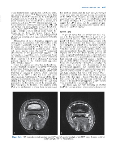
Figure 4.21. MRI images demonstrating a single large DDFT lesion (A; arrow) and multiple smaller DDFT lesions (B; arrow) at different
locations along the DDFT in the same horse.