
Page 488 - Adams and Stashak's Lameness in Horses, 7th Edition
P. 488
454 Chapter 4
VetBooks.ir
A B
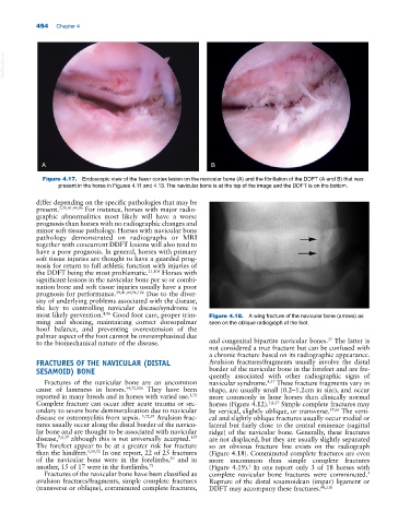
Figure 4.17. Endoscopic view of the flexor cortex lesion on the navicular bone (A) and the fibrillation of the DDFT (A and B) that was
present in the horse in Figures 4.11 and 4.13. The navicular bone is at the top of the image and the DDFT is on the bottom.
differ depending on the specific pathologies that may be
present. 3,39,41,44,96 For instance, horses with major radio-
graphic abnormalities most likely will have a worse
prognosis than horses with no radiographic changes and
minor soft tissue pathology. Horses with navicular bone
pathology demonstrated on radiographs or MRI
together with concurrent DDFT lesions will also tend to
have a poor prognosis. In general, horses with primary
soft tissue injuries are thought to have a guarded prog-
nosis for return to full athletic function with injuries of
the DDFT being the most problematic. 21,106 Horses with
significant lesions in the navicular bone per se or combi-
nation bone and soft tissue injuries usually have a poor
prognosis for performance. 39,41,44,96,106 Due to the diver-
sity of underlying problems associated with the disease,
the key to controlling navicular disease/syndrome is
most likely prevention. 4,96 Good foot care, proper trim- Figure 4.18. A wing fracture of the navicular bone (arrows) as
ming and shoeing, maintaining correct dorsopalmar seen on the oblique radiograph of the foot.
hoof balance, and preventing overextension of the
palmar aspect of the foot cannot be overemphasized due
37
to the biomechanical nature of the disease. and congenital bipartite navicular bones. The latter is
not considered a true fracture but can be confused with
a chronic fracture based on its radiographic appearance.
FRACTURES OF THE NAVICULAR (DISTAL Avulsion fractures/fragments usually involve the distal
SESAMOID) BONE border of the navicular bone in the forefeet and are fre-
quently associated with other radiographic signs of
Fractures of the navicular bone are an uncommon navicular syndrome. 8,37 These fracture fragments vary in
cause of lameness in horses. 54,72,116 They have been shape, are usually small (0.2–1.2 cm in size), and occur
reported in many breeds and in horses with varied use. 5,72 more commonly in lame horses than clinically normal
Complete fracture can occur after acute trauma or sec- horses (Figure 4.12). 7,8,37 Simple complete fractures may
ondary to severe bone demineralization due to navicular be vertical, slightly oblique, or transverse. 37,66 The verti-
disease or osteomyelitis from sepsis. 5,72,95 Avulsion frac- cal and slightly oblique fractures usually occur medial or
tures usually occur along the distal border of the navicu- lateral but fairly close to the central eminence (sagittal
lar bone and are thought to be associated with navicular ridge) of the navicular bone. Generally, these fractures
137
disease, 7,8,37 although this is not universally accepted. are not displaced, but they are usually slightly separated
The forefeet appear to be at a greater risk for fracture so an obvious fracture line exists on the radiograph
than the hindfeet. 5,59,72 In one report, 22 of 25 fractures (Figure 4.18). Comminuted complete fractures are even
of the navicular bone were in the forelimbs, and in more uncommon than simple complete fractures
59
another, 15 of 17 were in the forelimbs. 72 (Figure 4.19). In one report only 3 of 18 horses with
5
Fractures of the navicular bone have been classified as complete navicular bone fractures were comminuted.
5
avulsion fractures/fragments, simple complete fractures Rupture of the distal sesamoidean (impar) ligament or
(transverse or oblique), comminuted complete fractures, DDFT may accompany these fractures. 60,116