
Page 587 - Adams and Stashak's Lameness in Horses, 7th Edition
P. 587
Lameness of the Distal Limb 553
radiographic features of POD as focal radiopacities and/ Treatment
or radiolucencies in the palmar/plantar MC/MT III Treatment options are limited and range from rest,
VetBooks.ir phytosis (Figure 4.134). They reported a significant which allows for bone remodeling in the early stages of
condyles along with flattening of the condyle and osteo-
29
the POD, to systemic and intra‐articular anti‐inflamma-
improvement in sensitivity and specificity of diagnosis
of POD with plain radiographs following education of tory therapies. A period of 60‐ to 120‐day small pad-
reviewers on the common radiographic features of POD. dock turnout to allow for appropriate subchondral bone
The authors concluded that radiography is useful in remodeling was recommended by Tull et al., with 95%
95
later stages of disease but other imaging modalities are of horses diagnosed with POD returning to racing. If
29
required for early diagnosis of POD. Nuclear scintigra- progression of the disease continues, pain control is dif-
phy has been reported as a useful diagnostic tool for ficult and typically unrewarding in the majority of cases.
POD. In 2011, Trope et al. demonstrated that increased
IRU of the palmar/plantar MC/MT III condyles was the Prognosis
most common finding in the fetlock of Thoroughbred
racehorses and was associated with fewer starts and less Lameness associated with the early stages of POD
money earned than controls. The patterns of IRU with can resolve with appropriate rest from training allowing
93
nuclear scintigraphy are consistent with POD lesions the bone time to remodel and heal. As lesions progress
diagnosed on postmortem examination; however, there and cartilage damage occurs, progression to severe OA
has been no correlation with the degree of uptake and occurs, resulting in career‐ending lameness.
4
prognosis for return to racing. The use of MRI has
allowed for improved understanding of the relationship FETLOCK SUBCHONDRAL CYSTIC
between pathology and prognosis in cases of POD. In
2012, Powell described the appearance of POD lesions LESIONS (SCLS)
with standing low‐field MRI as regions of focal hyper- Fetlock subchondral bone cysts (SCLs) occur most
intensity along the margin of the joint surrounded commonly on the weight‐bearing surface of the MC/MT
by hypointense regions on STIR/T2* sequences. condyle and less commonly on the weight‐bearing sur-
Abnormalities were not detected radiographically in face of proximal P1. Cystic lesions of the distal metacar-
these horses. 73 pus/tarsus that open into the fetlock joint can occur in
On postmortem evaluation, lesions can range grossly young horses and are possibly considered part of the
from discoloration of the subchondral bone with mini- developmental osteochondrosis syndrome. 12
mal to no cartilage erosion to severe subchondral
bone discoloration and associated cartilage damage
(Figure 7.47). Histologically, common findings include Etiology
4
microfractures of the trabecular bone with thickening Some SCLs in the fetlock and other articular locations
and sclerosis of the adjacent bone, areas of osteocyte are considered part of the developmental orthopedic dis-
necrosis and eventual collapse, and degeneration of the ease complex that occur during growth and the conver-
overlying articular cartilage. 3 sion of cartilage to bone. However, some are thought to
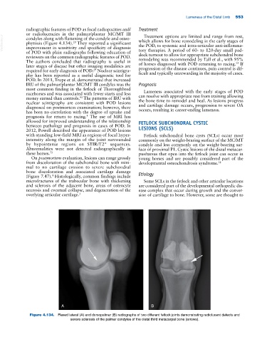
A B
Figure 4.134. Flexed lateral (A) and dorsopalmar (B) radiographs of two different fetlock joints demonstrating radiolucent defects and
severe sclerosis of the palmar condyles of the distal third metacarpal bone (arrows).