
Page 591 - Adams and Stashak's Lameness in Horses, 7th Edition
P. 591
Lameness of the Distal Limb 557
fractures of the proximal phalanx can accompany the articular surface and subchondral bone (Figure 4.138).
luxation. A varus (outward deviation of the cannon Radiographs are particularly important in young foals
VetBooks.ir (inward deviation of the cannon bone, outward devia- primary cause or a secondary contributing cause to the
to rule out the possibility of growth plate fractures as a
bone and inward deviation of the digit) or valgus
angular deformity.
tion of the digit) angular deformity is usually present
(Figure 4.137). Occasionally, the luxation will reduce
spontaneously, and the remaining evidence of its occur-
rence will be lameness, joint instability, and asymmetri- Treatment
cal fetlock swelling over the torn collateral ligament. Treatment of simple luxation of the fetlock can be
On palpation, the fetlock can usually be reduced and rewarding. In most cases the injury is limited to the sup-
re‐luxated without the degree of pain or evidence of porting soft tissues, and after the luxation is reduced
crepitation associated with fracture. More frequently, under anesthesia, good axial alignment can be main-
the swelling that is present is less than that observed tained by casting or splinting the limb until healing
with fracture, and the swelling is located selectively over occurs. Prior to applying the cast, needle drainage of the
the lateral or medial surface. Although the digital vascu- hematoma (if present) that overlies the ruptured collat-
lar supply is rarely compromised, it should be carefully eral ligament provides a better fit for the cast. Although
evaluated, particularly in open luxations. centesis of the hematoma and cast application can be
performed in the standing horse, general anesthesia and
lateral recumbency are preferred. Reduction of the luxa-
Diagnosis
tion is usually not difficult. A cast is applied that incor-
Generally, the diagnosis can be made by physical porates the foot and extends to just below the carpus or
examination alone. However, radiographs should be tarsus in the adult. Casts or splints are maintained for
taken to identify concurrent fractures or damage to the 6 weeks with stall rest. After cast or splint removal,
bandage support and limited exercise are recommended.
Swelling (thickening) will be noticed over the area of
collateral ligament rupture, and a cosmetic blemish will
remain. Additionally, some horses may have trouble
placing their heel on the ground when coming out of the
cast and may need a wedge shoe for a period of time.
A gradual return to exercise is recommended because
re‐luxation of the fetlock can occur if collateral ligament
healing is incomplete.
Success can be achieved without suture of the collat-
eral ligament, although there are several reports in the
literature on open repair. To repair the ligament, the end
is located after surgical incision, debrided, and sutured.
109
Alternatively, a polypropylene mesh has been substi-
tuted for the ruptured ligament. Arthroscopic removal
97
of the articular fractures should be elected if full athletic
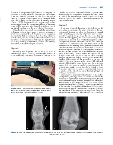
Figure 4.137. Image of closed subluxation of the left hind performance is a goal. This is not necessary for light rid-
fetlock prior to reduction and cast application. Note the lateral ing soundness if the fragments are small and from the
aspect of the distal third metatarsal bone (arrow). palmar/plantar eminence. Occasionally large avulsion
A B
Figure 4.138. (A) Standing and (B) stressed DP radiographs of a horse with subluxation of the fetlock with fragmentation of the collateral
ligament origin (arrow).