
Page 593 - Adams and Stashak's Lameness in Horses, 7th Edition
P. 593
Lameness of the Distal Limb 559
Perineural anesthesia, first of the foot and subse- Treatment
quently of the distal metacarpus or metatarsus, elimi- Treatment of affected horses is often dependent on
VetBooks.ir to the region of constriction. Direct anesthesia of the the stage of the disease, concurrent abnormalities, and
nates the lameness and locates the source of the lameness
intended use of the horse. Options usually include con-
tendon sheath is usually not necessary, but may be per-
formed if a fluid sample is being obtained for cytology servative medical therapy or desmopathy of the PAL
65
or if the sheath is injected as a conservative treatment. with or without tenoscopy. Adjunctive surgical proce-
However, anesthesia of the DFTS is fairly reliable for dures to simultaneously treat tendinitis, tendon core
documenting painful conditions within the sheath. In lesions, adhesions, and synovial proliferation are indi-
horses that respond dramatically to tendon sheath cated if the underlying conditions exist. Conservative
anesthesia, tenoscopy of the tendon sheath is often therapy is usually reserved for horses in the early stage
indicated. of the disease or those with primary PAL desmitis and
usually consists of oral and topical anti‐inflammatories,
intrasynovial treatment of the DFTS, controlled exer-
Diagnosis cise, and bandaging the limb. The most commonly used
surgical options for transection of the PAL is either a
The physical findings and the results of diagnostic anes- percutaneous approach or transection of the PAL con-
thesia provide a presumptive diagnosis. Ultrasonography currently at the time of tenoscopy. Tenoscopy of the
is indicated to identify the structures involved and assist DFTS is currently the preferred method to address syno-
with the selection of treatment. Normally the annular vial masses and tendon lesions and perform the annular
ligament is thin (less than 2 mm). One study found that ligament transection (Figure 4.139). Tenoscopy offers
measurement from the external skin surface to the internal the advantage of visualization of the sheath and syn-
annular ligament surface was the most reproducible index ovium and the tendons partially. Resection of adhesions,
of annular ligament thickening. Lesions identified at proliferative synovium, and tendon splitting can be per-
65
ultrasound vary and can include core tendon lesions or formed as desired. 33,101 If tenoscopy is not an option,
longitudinal tearing of the DDFT and SDFT, fibrosis, ten- then a percutaneous approach either above or in the
osynovitis and synovial proliferation, adhesions, and middle of the PAL can be performed. The PAL is tran-
thickening of the annular ligament. 30,65,96 Subcutaneous sected either with curved scissors or a curved blunt‐
fibrosis associated with PAL is common and should be tipped bistoury. Although many feel that surgical
differentiated from true thickening of the annular liga- transection is the ideal treatment, in one study of 71
ment itself. 65 horses with PAL injuries, the type of treatment did not
Radiographic evaluation of the fetlock region appear to influence the outcome. 65
should always be performed to evaluate possible osse-
ous involvement, particularly of the sesamoids. In
lesions associated with wounds or of traumatic origin, Prognosis
osteomyelitis or sequestrum of the sesamoid bones can Horses with PAL desmopathy alone that was not
be present concurrently with PAL constriction. This accompanied by abnormalities in the tendon (bowed
finding necessitates additional treatment and decreases tendon) had a good prognosis following surgical tran-
the prognosis for soundness or elimination of infec- section (84% returned to performance). 35,99 Other stud-
tion. In one study of 38 cases of annular ligament ies have reported a range of 50%–87% of horses with
constriction, 6 horses had proximal sesamoid bone desmitis of the PAL returning to athletic function. 55,65 If
abnormality, and 12 horses had bone enthesophytes at SDF tendinitis is present, the tendinitis rather than the
the attachment of the annular ligament (insertional constriction appears to limit the performance. In one
desmopathy). 87 study, of the 13 horses with SDF tendinitis together with
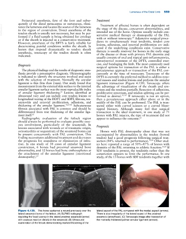
A B c
Figure 4.139. This horse sustained a wound previously over the lateral aspect of the PAL compared with the medial aspect (arrows).
lateral sesamoid bone of the fetlock. (A) DLPMO radiograph There is also irregularity of the lateral aspect of the proximal
depicting the focal lucency in the lateral proximal sesamoid (arrow) sesamoid (arrowhead). (C) Tenoscopic image after transection of
with osseous reaction distally in the sesamoid. (B) Ultrasound the markedly thickened palmar annular ligament (arrows).
examination of the fetlock demonstrating marked thickening of the