
Page 588 - Adams and Stashak's Lameness in Horses, 7th Edition
P. 588
554 Chapter 4
be associated with subchondral bone trauma and are Treatment
more common in adult horses. Injury to the articular Surgical debridement of metacarpal SCLs by an
VetBooks.ir impact may allow synovial fluid to enter the fissure and arthroscopic approach has historically been the prefer-
cartilage and underlying subchondral bone from high
able treatment if the diagnosis is made before significant
increase intraosseous pressure, contributing to cyst
42
development. Typically, the lameness and joint inflam- signs of OA have developed (Figure 4.135B). Medical
mation in these horses improve as the cyst matures. therapy with newer regenerative therapies or intra‐articu-
lar PSGAGs may also be helpful depending on the loca-
tion and characteristics of the SCLs. For SCLs that do
Clinical Signs not respond to conservative treatment or debridement,
The average age of horses with clinical signs in one osteostixis by extra‐articular approach or mosaic autol-
study was 18 months, although the SCL may develop ogous osteochondral grafting can be performed. 12
prior to the onset of clinical signs. Proximal P1 or
42
distal MC/MT SCLs can develop in horses of any age Prognosis
from trauma. Moderate lameness is often present
together with pain on fetlock flexion in most cases and The prognosis for return to performance appears to
fetlock joint effusion in approximately 50% of the be good for most horses with SCLs of the distal meta-
cases. However, the degree of lameness can vary con- carpus/tarsus. In one report, surgical treatment of third
siderably depending on the location and age of the SCL. metacarpal SCLs resulted in 80% of horses (12 of 15)
returning to their intended use. Follow‐up radio-
42
Diagnosis graphs to determine the degree of OA/degeneration of
the joint are helpful in determining the long‐term prog-
The diagnosis is usually made by radiographic evalu- nosis. Horses with trauma‐induced SCLs typically have
ation of the joint (Figure 4.135A). In the early develop- a more guarded prognosis for performance than those
ment of the SCL, it may not be apparent on radiographs, with developmental lesions regardless of their location.
and follow‐up radiographic examination is recom-
mended if clinical signs that refer to the joint persist.
Nuclear scintigraphy can identify a focal area of TRAUMATIC RUPTURE OF THE SUSPENSORY
increased uptake in the bone as the SCL is developing APPARATUS
and for several months after it has formed. Chronic
SCLs may not have significant bone turnover above the Traumatic rupture of the suspensory apparatus with
surrounding condyle and may not be detectable with or without fractures of both proximal sesamoid bones is
scintigraphy. Advanced imaging (CT or MRI) is often a common cause of acute breakdown in the racing
helpful to document the extent of the SCL and any sec- Thoroughbred and often results in humane destruction
ondary articular pathology that may be present in horses of the animal. 14,40,46,77 Proximal luxations of the sesa-
with trauma‐induced lesions. moid bone without fracture can also occur with trau-
A B
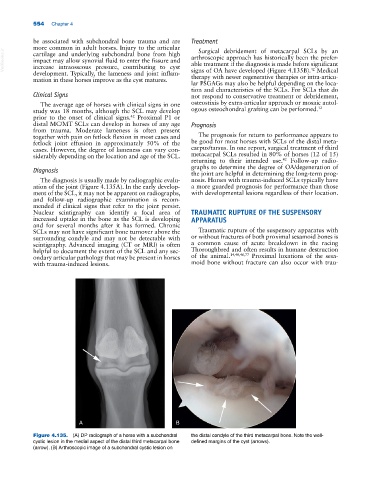
Figure 4.135. (A) DP radiograph of a horse with a subchondral the distal condyle of the third metacarpal bone. Note the well‐
cystic lesion in the medial aspect of the distal third metacarpal bone defined margins of the cyst (arrows).
(arrow). (B) Arthroscopic image of a subchondral cystic lesion on