
Page 598 - Adams and Stashak's Lameness in Horses, 7th Edition
P. 598
564 Chapter 4
taken at this time are usually negative; however, frac-
tures are occasionally observed as well as a minimal
VetBooks.ir ably develops as a result of acute disease that is unre-
amount of superficial cortical osteolysis.
Subacute to chronic dorsal metacarpal disease invari-
sponsive to therapy or has gone unrecognized. It is most
frequently seen in horses 2–4 years of age. Only mild
86
degrees of gait deficit may be noticed at exercise. On
palpation, varying degrees of pain may be elicited, but a
more obvious enlargement is palpable on the dorsome-
dial cortex (Figure 4.140). The pain response is typically
more profound after strenuous exercise, and the left
limb is usually more severely affected. Periosteal new
bone formation is usually observed on radiographic
examination (Figures 4.140 and 4.141).
Dorsal metacarpal bone failure or stress fractures
usually occur in the dorsolateral cortex, although frac-
tures can occur in the dorsal or dorsomedial cortex. It is
usually observed in older horses, 3–5 years of age, that
have a history of bucked shins. As with dorsal metacar-
pal disease, the lameness may not be prominent while
the horse is rested. However, it is usually prominent
after strenuous exercise. On palpation, a rather discrete
painful area can be palpated on the dorsolateral, or less
commonly dorsomedial, surface of the left third meta-
carpal (cannon) bone at the junction of its middle and
A B
distal third. Only rarely is the right third metacarpal
bone involved. Radiographs usually point to a cortical
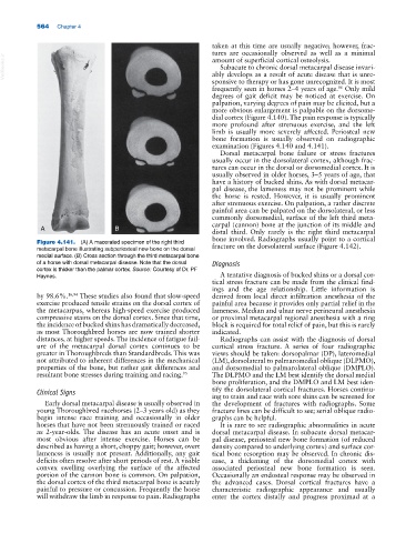
Figure 4.141. (A) A macerated specimen of the right third fracture on the dorsolateral surface (Figure 4.142).
metacarpal bone illustrating subperiosteal new bone on the dorsal
medial surface. (B) Cross section through the third metacarpal bone
of a horse with dorsal metacarpal disease. Note that the dorsal Diagnosis
cortex is thicker than the palmar cortex. Source: Courtesy of Dr. PF
Haynes. A tentative diagnosis of bucked shins or a dorsal cor-
tical stress fracture can be made from the clinical find-
ings and the age relationship. Little information is
by 98.6%. 16,94 These studies also found that slow‐speed derived from local direct infiltration anesthesia of the
exercise produced tensile strains on the dorsal cortex of painful area because it provides only partial relief in the
the metacarpus, whereas high‐speed exercise produced lameness. Median and ulnar nerve perineural anesthesia
compressive stains on the dorsal cortex. Since that time, or proximal metacarpal regional anesthesia with a ring
the incidence of bucked shins has dramatically decreased, block is required for total relief of pain, but this is rarely
as most Thoroughbred horses are now trained shorter indicated.
distances, at higher speeds. The incidence of fatigue fail- Radiographs can assist with the diagnosis of dorsal
ure of the metacarpal dorsal cortex continues to be cortical stress fracture. A series of four radiographic
greater in Thoroughbreds than Standardbreds. This was views should be taken: dorsopalmar (DP), lateromedial
not attributed to inherent differences in the mechanical (LM), dorsolateral to palmaromedial oblique (DLPMO),
properties of the bone, but rather gait differences and and dorsomedial to palmarolateral oblique (DMPLO).
resultant bone stresses during training and racing. 95 The DLPMO and the LM best identify the dorsal medial
bone proliferation, and the DMPLO and LM best iden-
Clinical Signs tify the dorsolateral cortical fractures. Horses continu-
ing to train and race with sore shins can be screened for
Early dorsal metacarpal disease is usually observed in the development of fractures with radiographs. Some
young Thoroughbred racehorses (2–3 years old) as they fracture lines can be difficult to see; serial oblique radio-
begin intense race training and occasionally in older graphs can be helpful.
horses that have not been strenuously trained or raced It is rare to see radiographic abnormalities in acute
as 2‐year‐olds. The disease has an acute onset and is dorsal metacarpal disease. In subacute dorsal metacar-
most obvious after intense exercise. Horses can be pal disease, periosteal new bone formation (of reduced
described as having a short, choppy gait; however, overt density compared to underlying cortex) and surface cor-
lameness is usually not present. Additionally, any gait tical bone resorption may be observed. In chronic dis-
deficits often resolve after short periods of rest. A visible ease, a thickening of the dorsomedial cortex with
convex swelling overlying the surface of the affected associated periosteal new bone formation is seen.
portion of the cannon bone is common. On palpation, Occasionally an endosteal response may be observed in
the dorsal cortex of the third metacarpal bone is acutely the advanced cases. Dorsal cortical fractures have a
painful to pressure or concussion. Frequently the horse characteristic radiographic appearance and usually
will withdraw the limb in response to pain. Radiographs enter the cortex distally and progress proximad at a