
Page 599 - Adams and Stashak's Lameness in Horses, 7th Edition
P. 599
Lameness of the Distal Limb 565
still in training. Racehorses may have bone scintigrams
performed to determine whether training should con-
VetBooks.ir fracture. In a review of 121 bone scintigrams of racing
tinue or to identify focal uptake indicative of impending
Thoroughbreds with clinical history of dorsal metacar-
pal disease, horses without fractures (bucked shins) had
a mild to moderate diffuse uptake of radioisotope,
unlike the focal intense uptake typical of dorsal cortical
fracture. Chronic fractures are less focal and intense
because of surrounding bone remodeling; however, they
still appear relatively focal compared to early disease.
Standing robotic cone‐beam computed tomography
(CT) might be useful in defining difficult‐to‐see frac-
tures. Use of quantitative CT to measure bone mineral
density and three‐dimensional morphologic changes in
the metacarpus may provide improvements in identify-
ing risk factors or imminence of fracture.
Treatment
There is no specific medical treatment for dorsal met-
A acarpal disease, necessitating that horses be laid up or
put on a convalescent exercise program to provide time
for the early acute changes to subside. Many horses with
acute bucked shins can continue to train after 5–10 days
of rest and anti‐inflammatory analgesics. Hand walking,
ponying, cold water hosing, and bandaging should con-
tinue until the dorsal cortex can be palpated without
eliciting pain. Speed and distance are introduced slowly,
with constant monitoring of dorsal cortical pain.
Initially, daily galloping distance is reduced to 50%. An
overall modification of the training program with less
galloping miles and more short‐distance breezing may
be necessary. The overall goal is to gradually increase
the stress to the dorsal surface of the metacarpal bone at
such a rate such that this surface can model according to
compressive demands without producing structural
B damage.
Subacute and chronic dorsal metacarpal disease can
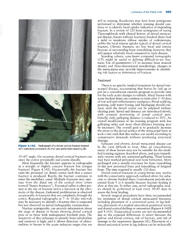
Figure 4.142. Radiograph of a dorsal cortical fracture treated be the most difficult to treat. After an exacerbation,
with osteostixis procedure (A) that was performed standing (B). many of these horses may not be suitable for the modi-
fied training regimen described above, and pain immedi-
35–45° angle. On occasion, dorsal cortical fractures can ately returns with any sustained galloping. These horses
enter the cortex proximally and course distally. may have marked periosteal new bone formation. More
Most frequently the fracture appears in radiographs prolonged rest is usually necessary for bone remodeling
as a straight or slightly concave fracture line (tongue of this new periosteal bone and remodeling of fatigued
fracture) (Figure 4.142). Occasionally, the fracture line bone. The time required is usually 110 days.
exits the proximal (or distal) cortex such that a saucer Dorsal cortical fractures in young horses may resolve
fracture is produced. Rarely, the fracture continues to with the conservative approach outlined above for suba-
enter the medullary canal. Multiple fractures may ema- cute or chronic bucked shins. Convalescent periods may
nate from the distal site of the cortical entry (often extend from 4 to 6 months because fracture healing is
termed “fissure fractures”). Periosteal callus is often pre- slow at this site. In either case, serial radiographic stud-
sent at the site of fracture and is a function of the chro- ies should be performed at least every 30–45 days to
nicity of the disease. Endosteal proliferation is observed assess the bone healing.
occasionally in fractures that are completely through the Several surgical procedures have been recommended
cortex. Repeated radiographs at 7‐ to 10‐day intervals for treatment of dorsal cortical metacarpal fractures,
may be necessary to identify a fracture that is suspected including placement of a unicortical screw in lag fash-
but not observed on initial radiographic examination. ion, placement of a neutral unicortical positional screw,
Nuclear scintigraphy can provide information about and dorsal cortical drilling or osteostixis (Figures 4.142B
the stage of disease in horses showing dorsal cortical and 4.143). Transcortical screws are not recommended
pain or in those with undiagnosed forelimb pain. The due to the expected differences in strain between the
sensitivity of this technique to identify bone metabolism palmar and dorsal cortices, risk of fracture, and risk of
and turnover is high, and it allows detection of abnor- damage to the suspensory ligament (SL). Placement of a
malities in horses in the acute–subacute stages that are dorsal unicortical screw in lag fashion can be technically