
Page 600 - Adams and Stashak's Lameness in Horses, 7th Edition
P. 600
566 Chapter 4
Osteostixis can be performed under general anesthe-
sia or in the standing horse (Figure 4.142B). Holes (5–7)
VetBooks.ir mond pattern through the dorsal cortex into the mar-
are made with a small drill bit (2.0–2.5 mm) in a dia-
A
row cavity. This procedure can be combined with
unicortical screw application. Success for return to rac-
ing is similar (greater than 80%) to that recently reported
for screw fixation, and a quicker return to racing (4–6
months) has been reported. Clustered drill holes act as
57
a stress concentrator and significantly decrease the stress
required for metacarpal failure in cadaver limbs, but
B
catastrophic failure through the drill holes is not a
reported complication of the procedure in vivo. 129,130
Other adjunctive treatments have been recommended
with or without surgical treatment, including electrical
stimulation at the fracture, extracorporeal shockwave
therapy, injection of osteogenic substances (sodium
oleate), intralesional injection of steroids, thermocau-
tery (pin firing), chemical vesication (blistering), needle
C drainage of the hematoma, and cryotherapy (point
freezing). These treatments have met with varying
degrees of success, and no controlled studies have been
performed. In all cases, no matter what the treatment, an
adequate period of rest combined with a controlled
exercise program is required.
In one study of Thoroughbred racehorses, distinct
training strategies were used at various stables, and the
allocation of exercise to breezing (15 m/s), galloping
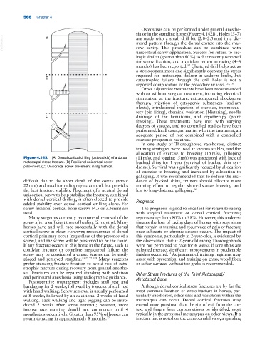
Figure 4.143. (A) Dorsal cortical drilling (osteostixis) of a dorsal (11 m/s), and jogging (5 m/s) was associated with lack of
metacarpal stress fracture. (B) Positional unicortical screw bucked shins for 1 year (survival of bucked shin syn-
placement. (C) Unicortical screw placement in lag fashion. drome). Survival was significantly reduced by allocation
of exercise to breezing and increased by allocation to
galloping. It was recommended that to reduce the inci-
difficult due to the short depth of the cortex (about dence of bucked shins, trainers should allocate more
22 mm) and need for radiographic control, but provides training effort to regular short‐distance breezing and
the best fracture stability. Placement of a neutral dorsal less to long‐distance galloping. 16
unicortical screw to help stabilize the fracture, combined
with dorsal cortical drilling, is often elected to provide Prognosis
added stability over dorsal cortical drilling alone. For
screw fixation, cortical bone screws (4.5 or 3.5 mm) are The prognosis is good to excellent for return to racing
used. with surgical treatment of dorsal cortical fractures;
Many surgeons currently recommend removal of the reports range from 80% to 98%. However, this underes-
screw after a sufficient time of healing (2 months). Many timates the loss of racing days of horses with sore shins
horses have and will race successfully with the dorsal that remain in training and recurrence of pain or fracture
cortical screw in place. However, reoccurrence of dorsal once subacute or chronic disease occurs. The impact of
cortical pain may occur (regardless of the presence of a this syndrome, particularly in 2‐year‐olds, is evidenced by
screw), and the screw will be presumed to be the cause. the observation that if 2‐year‐old racing Thoroughbreds
If any fracture occurs in this horse in the future, such as were not permitted to race for 6 weeks if sore shins are
condylar fracture or complete metacarpal failure, the palpated prerace, significant improvements in predictable
screw may be considered a cause. Screws can be easily finishes occurred. Adjustment of training regimens may
55
placed and removed standing. 23,27,57,129 Many surgeons assist with prevention, and training on grass, wood fiber,
prefer standing fracture fixation to avoid risk of cata- or softer surfaces without toe grabs is recommended.
strophic fracture during recovery from general anesthe-
sia. Fractures can be repaired standing with sedation Other Stress Fractures of the Third Metacarpal/
and perineural anesthesia using radiographic guidance. Metatarsal Bone
Postoperative management includes stall rest and
bandaging for 2 weeks, followed by 6 weeks of stall rest Although dorsal cortical stress fractures are by far the
with hand walking. Screw removal is usually performed most common location of stress fracture in horses, par-
at 8 weeks, followed by an additional 2 weeks of hand ticularly racehorses, other sites and variations within the
walking. Tack walking and light jogging can be intro- metacarpus can occur. Dorsal cortical fractures may
duced 2 weeks after screw removal; however, more extend more proximal than the site of exit from the cor-
intense race training should not commence until 4 tex, and fissure lines can sometimes be identified, most
months postoperatively. Greater than 95% of horses can typically in the proximal metacarpus on other views. If a
return to racing in approximately 8 months. 27 fracture line is noted on the craniocaudal view, a spiraling