
Page 603 - Adams and Stashak's Lameness in Horses, 7th Edition
P. 603
Lameness of the Distal Limb 569
VetBooks.ir
A B C
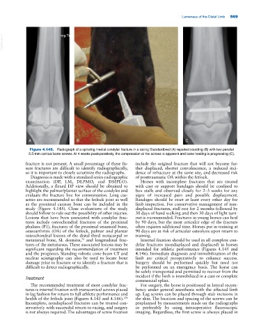
Figure 4.145. Radiograph of a spiraling medial condylar fracture in a racing Standardbred (A) repaired standing (B) with two parallel
5.5‐mm cortical bone screws. At 4 weeks postoperatively, the compression at the screws is apparent and bone healing is progressing (C).
fracture is not present. A small percentage of these fis- include the original fracture that will not become fur-
sure fractures are difficult to identify radiographically, ther displaced, shorter convalescence, a reduced inci-
so it is important to closely scrutinize the radiographs. dence of refracture at the same site, and decreased risk
Diagnosis is made with a standard series radiographic of posttraumatic OA within the fetlock.
examination (DP, LM, DLPMO, and DMPLO). Horses with incomplete fractures that are treated
Additionally, a flexed DP view should be obtained to with cast or support bandages should be confined to
highlight the palmar/plantar surface of the condyles and box stalls and observed closely for 2–3 weeks for any
evaluate the fracture line for comminution. Long cas- signs of increased pain and possible displacement.
settes are recommended so that the fetlock joint as well Bandages should be reset at least every other day for
as the proximal cannon bone can be included in the limb inspection. For conservative management of non-
study (Figure 4.145). Close evaluations of the study displaced fractures, stall rest for 2 months followed by
should follow to rule out the possibility of other injuries. 30 days of hand walking and then 30 days of light turn-
Lesions that have been associated with condylar frac- out is recommended. Fractures in young horses can heal
tures include osteochondral fractures of the proximal by 90 days, but the most articular edge of the fracture
phalanx (P1), fractures of the proximal sesamoid bone, often requires additional time. Horses put in training at
osteoarthritis (OA) of the fetlock, palmar and plantar 90 days are at risk of articular osteolysis upon return to
osteochondral lesions of the distal third metacarpal or training.
metatarsal bone, SL desmitis, and longitudinal frac- Internal fixation should be used in all complete con-
73
tures of the metatarsus. These associated lesions may be dylar fractures (nondisplaced and displaced) in horses
significant regarding the recommendation of treatment intended for athletic performance (Figures 4.145 and
and the prognosis. Standing robotic cone‐beam CT and 4.146). Immediate diagnosis and immobilization of the
nuclear scintigraphy can also be used to locate bone limb are critical preoperatively to enhance success.
damage prior to fracture or to identify a fracture that is Surgery should be performed quickly but need not
difficult to detect radiographically. be performed on an emergency basis. The horse can
be safely transported and permitted to recover from the
Treatment incident if the limb is immobilized in a cast or complete
commercial splint.
The recommended treatment of most condylar frac- For surgery, the horse is positioned in lateral recum-
tures is internal fixation with transcortical screws placed bency under general anesthesia with the affected limb
in lag fashion for return to full athletic performance and up. Lag screws can be placed through stab incisions in
126
health of the fetlock joint (Figures 4.145 and 4.146). the skin. The location and spacing of the screws can be
Incomplete, nondisplaced fractures can be treated con- preplanned by measurements made on the radiographs
servatively with successful return to racing, and surgery or preferably by using intraoperative fluoroscopic
is not always required. The advantages of screw fixation imaging. Regardless, the first screw is always placed in