
Page 606 - Adams and Stashak's Lameness in Horses, 7th Edition
P. 606
572 Chapter 4
stages, 2–3 months apart. After plate removal, stall rest
is continued for an additional 2 months and then exer-
VetBooks.ir Prognosis
cise is slowly initiated.
The prognosis for successful treatment of third meta-
carpal and metatarsal bone fractures depends on multi-
ple factors. In general, transverse, slightly oblique, and
slightly comminuted fractures in the mid‐cannon bone
region in foals under 7 months of age have a good to
excellent prognosis with internal fixation. Older horses
with similar fractures have a more guarded prognosis
due to their size and the risk of supporting limb laminitis,
but in general have a fair to good prognosis. Older horses
with open, comminuted, or articular fractures have a
guarded to poor prognosis for recovery. Unfortunately,
older horses have a greater risk of comminuted open
fractures that involve the nutrient foramen. 84
METACARPAL/METATARSAL EXOSTOSIS
(SPLINTS)
Exostoses of the second and fourth metacarpal/meta-
tarsal bones, or “splints,” are a condition most commonly
diagnosed in young, immature horses but can be seen in
older horses. The condition is thought to be associated
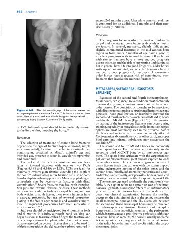
Figure 4.147. This oblique radiograph of the tarsus revealed an with direct trauma causing subperiosteal hemorrhage and
incomplete proximal metatarsal fracture. This fracture occurred from periosteal bone proliferation or instability between the
an accident at a jump and was initially thought to be a proximal second and fourth metacarpal/metatarsal (MC/MT) bones
suspensory injury. Source: Courtesy of Dr. Ty Wallis. and the third MC/MT bone (Figure 4.150). Inflammation
or tearing of the interosseous ligament can occur during
or PVC full‐limb splint should be immediately secured training, especially in musculoskeletally immature horses.
to the limb without moving the horse. 13 Splints are most commonly seen in the proximal half of
the bones and metacarpal II is most commonly affected.
Conformation abnormalities such as offset carpi, improper
Treatment
hoof care, and mineral imbalances may exacerbate the
The selection of treatment of cannon bone fractures condition. 108
depends on the type of fracture (open vs. closed, simple The second and fourth MC/MT bones are commonly
vs. comminuted), location of the fracture (articular vs. called splint bones. Each is attached intimately to the
nonarticular, proximal vs. distal), animal’s age and respective third MC/MT bone by an interosseous liga-
intended use, presence of wounds, vascular compromise, ment. The splint bones articulate with the carpometacar-
and economics. pal joint or tarsometatarsal joint and are exposed to loads
The preferred treatment for most cannon bone frac- on weight‐bearing. The interosseous ligament consists of
tures is internal fixation with one or two DCPs dense fibrous tissue that can tear with the strains applied
(Figures 4.148 and 4.149) or LCPs. LCPs are ideal for during independent motion of the splint bones and the
minimally invasive plate fixation extending the length of cannon bone. Initially, inflammatory periostitis and desmi-
the bone. Individual lag screw fixation can also be com- tis develop. Subsequently, new periosteal bone is produced,
64
bined with plates where appropriate. Metacarpophalangeal creating the characteristic profile of a splint (Figure 4.150).
arthrodesis may be necessary in fractures with distal The terminology used to identify the condition is var-
comminution. Severe fractures may heal with transfixa- iable. A true splint refers to a sprain or tear of the inter-
19
tion pins and external fixators or casts. These methods osseous ligament. Blind splint refers to an inflammatory
are more successful in foals with rapid healing and low process of the interosseous ligament that is difficult to
body weight. In adult horses, these severe fractures have detect on physical examination because the swelling
a guarded prognosis, but use of external coaptation, occurs on the axial (inner) side of the splint, between the
plating in the face of open wounds and vascular compro- small metacarpal bone and the SL. Osteolysis between
mise, or sequential procedures have been successful in the second and third metacarpal bones may be observed
rare instances. 14,64,76 on radiographic examination. Periostitis of the splint
Stall rest should be anticipated for 4 months in foals bones results from superficial trauma to the periosteum,
and 6 months in adults, although hand walking can which, in turn, causes a proliferative periostitis. Although
begin as soon as fracture callus bridges the fracture and a residual blemish remains, the horse is usually not lame.
other complications of implant failure have not occurred A knee splint is the enlargement of the proximal portion
or have arrested. In general, horses that are to be used in of the splint bone that may lead to OA within the carpo-
athletic competition should have their plates removed in metacarpal joint.