
Page 609 - Adams and Stashak's Lameness in Horses, 7th Edition
P. 609
Lameness of the Distal Limb 575
Treatment Intralesional corticosteroid can reduce inflammation
and may help prevent excessive bone growth. Corticosteroid
Treatment for splints includes anti‐inflammatory
VetBooks.ir agents and rest for the acute phase and occasionally sur- bandage. In this case the horse is generally rested longer
therapy should be accompanied by counterpressure
gery in the more chronic stages. Counterirritation is still
than 30 days and should not resume training as rapidly
practiced for the chronic phases but is of unknown sig-
nificance. Other treatments include extracorporeal as when counterirritation is used. However, the swelling
may be considerably less. It is also true that splints will
shockwave therapy, icing, topical diclofenac liposomal heal without therapy, provided adequate rest is given.
108
cream (Surpass), acupuncture, and massage. If the splint results from interference, splint or shin boots
Inflammation and swelling are the hallmark of this (guards) may help prevent further trauma. If the horse
disease in the acute phase. The administration of non- interferes because of improper trimming and shoeing,
steroidal anti‐inflammatory drugs (NSAIDs) coupled this should be corrected.
with the application of hypothermia and pressure/sup- If the proliferative bone is excessive, surgery may be
port wraps appears to be most beneficial to decrease the indicated in a very small percentage of the cases
heat, pain, and swelling. Hypothermia can be attained (Figure 4.151). Surgery to remove exostoses for medical
with ice or ice/water packs or whirlpool boots. They or cosmetic reasons has resulted in fair to good success.
should be applied for 30 minutes, 2–3 times a day for at In one study, 15 exostoses removed for cosmetics or
least 2–3 days. Some recommend hand massage for 10 lameness resulted in minimal recurrence of the bone
minutes after each treatment, after which a support proliferation. In a larger study of 95 Standardbreds, the
5
bandage is applied. Affected horses should be confined splint bones were amputated to remove excessive bone
to a stall for at least 30–45 days, and hand‐walking callus. In horses with proximal splint bone removal in
exercise for 15–20 minutes twice a day should be begun which the proximal portion was stabilized with screws
after the acute inflammation subsides. 108 or bone plates, horses were still limited in performance
at 12 weeks. Horses in which a subperiosteal removal of
the exostosis was performed were sound 12 weeks post-
141
operatively. It is recommended to retain the splint
bone lever arm by reflecting the periosteum to prevent
Figure 4.151. This large exostosis of the medial splint (arrows)
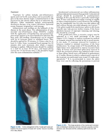
Figure 4.150. Visible enlargement of the medial splint area just was contributing to lameness and was removed surgically. Just the
distal to the carpus typical of horses with “splints.” Source: Courtesy exostosis was removed and the underlying splint bone was left
of Dr. Gary Baxter. intact.