
Page 610 - Adams and Stashak's Lameness in Horses, 7th Edition
P. 610
576 Chapter 4
formation of excessive new bone and associated irrita- the distal third. 3,17,31,32,35,136 In most cases fractures
tion of the SL. In some cases, it is necessary to surgically located at the distal third are simple fractures
VetBooks.ir of the SL or the carpal joint or one that is so large that and proximal portion, which are often complicated by
(Figure 4.152), in contrast to fractures of the middle
remove a bony exostosis that interferes with the action
comminution, osteomyelitis, and bone sequestration
the opposite foot hits it repeatedly. These require care in
dissection of the proximal structures, including liga- (Figure 4.153). 10,31,58,101
ments of the palmar carpal support. If the bone growth Fractures of the distal part of a small metacarpal or
has been caused by trauma from interference, the sur- metatarsal bone usually occur in older horses (5–7 years
gery will not be successful unless corrective shoeing or of age) and only rarely occur in horses under 2 years of
use of splint boots will stop the interference. age. This is thought to occur as a result of decreased
17
pliability in the interosseous ligament and more strenu-
17
Prognosis ous training programs in older horses. In contrast,
younger horses tend to sustain damage to the interos-
Prognosis is good to excellent for soundness except seous ligament supporting the small metacarpal bones,
for those in which the exostosis is large and encroaches resulting in the condition referred to as splints. The
on the SL or the carpal joint. Chronic recurring lame- forelimbs are more frequently involved than the
ness can occur in horses that are not rested long enough, hindlimbs; the fourth metacarpal bone in the left fore-
which can be 5–6 months. Surgery to remove the excess limb is affected more commonly than the hindlimb. 17,136
bone callus can successfully alleviate lameness, and The relationship between SL desmitis, sesamoiditis,
recurrence does not occur in most cases. Surgery may and fetlock OA is more than casual. It appears that the
speed up the return to athletic soundness and improve enlarged fibrotic SL decreases the absorptive capacity
the cosmetic blemish. of the fetlock and creates a space‐occupying mass that
may lead to fracture of the small metacarpal/metatar-
sal bones followed by further displacement of the
FRACTURES OF THE SMALL METACARPAL AND fracture fragment.
METATARSAL (SPLINT) BONES Fractures of the proximal half of the small metacar-
pal or metatarsal bone are often comminuted. Open
Fractures of the small metacarpal and metatarsal fractures can often be complicated by osteomyelitis with
bones (splint bones) can occur anywhere along or without sequestrum formation. Fractures of the prox-
their length, but they are most commonly located at imal lateral splint bones are usually a result of direct
trauma (Figure 4.153). 10,58,101
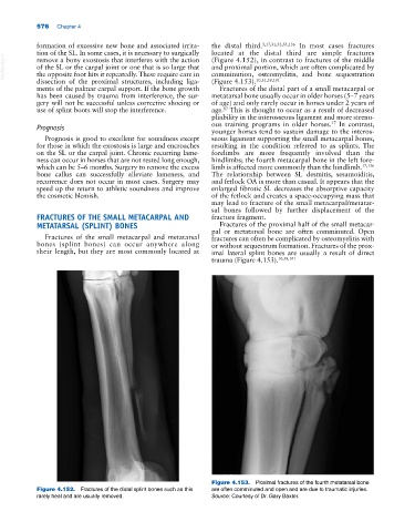
Figure 4.153. Proximal fractures of the fourth metatarsal bone
Figure 4.152. Fractures of the distal splint bones such as this are often comminuted and open and are due to traumatic injuries.
rarely heal and are usually removed. Source: Courtesy of Dr. Gary Baxter.