
Page 615 - Adams and Stashak's Lameness in Horses, 7th Edition
P. 615
Lameness of the Distal Limb 581
occurs in Standardbreds, hunters and jumpers, and polo distal to the carpometacarpal or tarsometatarsal joints
ponies, but other breeds are affected as well. 137 (Figure 4.160). Overloading of the SL may cause trauma
VetBooks.ir occurs most frequently in racehorses, specifically seen more commonly in sport horses. Hyperextension of
to any portion of the ligament, but origin injuries are
Injury to the body of the SL is less common and
the carpus/tarsus in conjunction with severe overexten-
Standardbred racehorses. The body of the ligament also
is usually involved in cases of degenerative suspensory sion of the fetlock joint has been proposed to cause
ligament desmitis (DSLD). In general, lesions of the proximal lesions. In general, the more severe the trauma,
56
body of the suspensory have a more guarded prognosis the more severe the ligamentous lesion. Working horses
for return to full athletic work than proximal injury or in deep, soft arenas or in eventing where there is exces-
injury of the branches of the suspensory. 143 sive rotational movement of the limbs may increase the
Injury to the SL branches occurs most commonly in risk of injuries. Lesions in the body or branches of the
Standardbred racehorses or jumping horses. “Bowing” or suspensory also occur in sport horses worked in soft
“springing” of the distal ends of the splint bones and frac- ground. Therefore, soft tissue injuries, including suspen-
ture of the distal splint bones is associated with desmitis sory injuries, occur more frequently in Europe where
of the branches of the suspensory. This finding is consid- horses race and train on turf. Lesions within the branches
ered a result of the enlargement of the branches physically are also associated with fetlock lameness and suggest
forcing the ends of the splint bones abaxially. This predis- that high rotary motion of the fetlock may predispose to
poses the splint bones to fail or fracture at their thinnest suspensory branch injury as may occur in racehorses
point. These fractures do not heal spontaneously because and animals with dropped fetlock conformation.
the splint bones are under tension. Lameness is often due
to the suspensory problem and not the fracture.
Clinical Signs
Most horses with PSD present with a history of inter-
Etiology
mittent lameness of several days’ or week’s duration that
The SL is primarily made up of dense white fibrous is exacerbated by resumed exercise. The onset may be
connective tissue. Proximally, the body of the SL sepa- acute or insidious. Generally, heat and swelling are pal-
rates and attaches into two palmar depressions just pable on the proximal aspect of the limb only in acute
cases. In chronic intermittent cases, physical findings are
less obvious or may not be present to assist with a diag-
nosis. However, slight proximal swelling may be felt on
the medial side between the SL and deep digital flexor
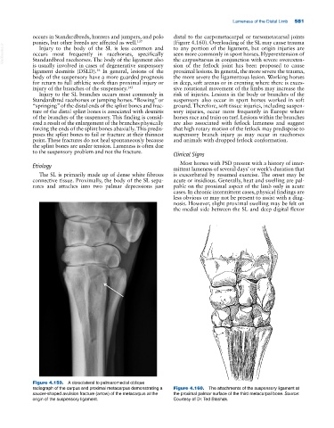
Figure 4.159. A dorsolateral to palmaromedial oblique
radiograph of the carpus and proximal metacarpus demonstrating a Figure 4.160. The attachments of the suspensory ligament at
saucer‐shaped avulsion fracture (arrow) of the metacarpus at the the proximal palmar surface of the third metacarpal bone. Source:
origin of the suspensory ligament. Courtesy of Dr. Ted Stashak.