
Page 620 - Adams and Stashak's Lameness in Horses, 7th Edition
P. 620
586 Chapter 4
VetBooks.ir
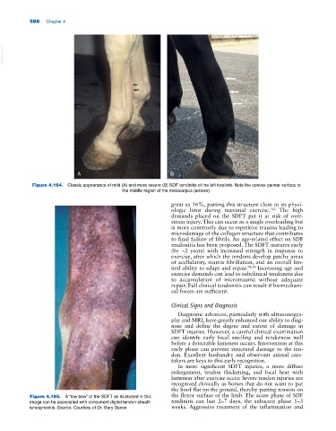
A B
Figure 4.164. Classic appearance of mild (A) and more severe (B) SDF tendinitis of the left forelimb. Note the convex palmar surface to
the middle region of the metacarpus (arrows).
great as 16%, putting this structure close to its physi-
ologic limit during maximal exercise. 142 The high
demands placed on the SDFT put it at risk of over-
strain injury. This can occur as a single overloading but
is more commonly due to repetitive trauma leading to
microdamage of the collagen structure that contributes
to final failure of fibrils. An age‐related effect on SDF
tendonitis has been proposed. The SDFT matures early
(by ~2 years) with increased strength in response to
exercise, after which the tendons develop patchy areas
of acellularity, matrix fibrillation, and an overall lim-
ited ability to adapt and repair. 98,99 Increasing age and
exercise demands can lead to subclinical tendonitis due
to accumulation of microtrauma without adequate
repair. Full clinical tendonitis can result if biomechani-
cal forces are sufficient.
Clinical Signs and Diagnosis
Diagnostic advances, particularly with ultrasonogra-
phy and MRI, have greatly enhanced our ability to diag-
nose and define the degree and extent of damage in
SDFT injuries. However, a careful clinical examination
can identify early focal swelling and tenderness well
before a detectable lameness occurs. Intervention at this
early phase can prevent structural damage to the ten-
don. Excellent husbandry and observant animal care-
takers are keys to this early recognition.
In more significant SDFT injuries, a more diffuse
enlargement, tendon thickening, and local heat with
lameness after exercise occur. Severe tendon injuries are
recognized clinically as horses that do not want to put
the hoof flat on the ground, thereby putting tension on
Figure 4.165. A “low bow” of the SDFT as illustrated in this the flexor surface of the limb. The acute phase of SDF
image can be associated with concurrent digital tendon sheath tendinitis can last 2–7 days, the subacute phase 1–3
tenosynovitis. Source: Courtesy of Dr. Gary Baxter. weeks. Aggressive treatment of the inflammation and