
Page 625 - Adams and Stashak's Lameness in Horses, 7th Edition
P. 625
Lameness of the Distal Limb 591
tudinal tears occur within the sheath, and effusion
should direct the clinician to further investigation of the
VetBooks.ir tendon sheath tenosynovitis.
digital tendon sheath. See the section on digital flexor
Treatment
Treatment of DDF tendinitis is similar to that of SDF
tendinitis in terms of physical therapy, systemic medical
management, and convalescence. DDF tendinitis may be
treated surgically with distal accessory desmotomy for
the same reasons that SDF tendinitis may be treated with
proximal accessory desmotomy to relieve tension on the
11
DDFT. Literature supporting this treatment is limited,
partly due to the rarity of the condition. Injuries to the
DDFT within the sheath are frequently treated as a DDF
tenosynovitis, in addition to treatment of the tendon
injury. See the digital tenosynovitis in the fetlock section.
Prognosis
The prognosis for DDF tendinitis is guarded, depend-
ing on severity. In one study of 24 cases, only 7 returned
139
to athletic use.
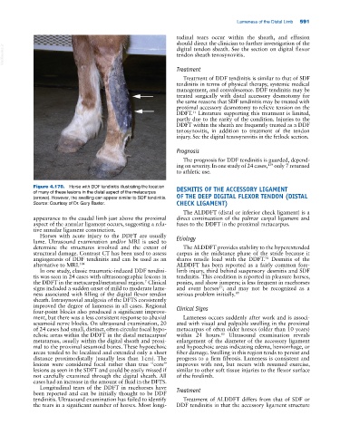
Figure 4.170. Horse with DDF tendinitis illustrating the location DESMITIS OF THE ACCESSORY LIGAMENT
of many of these lesions in the distal aspect of the metacarpus
(arrows). However, the swelling can appear similar to SDF tendinitis. OF THE DEEP DIGITAL FLEXOR TENDON (DISTAL
Source: Courtesy of Dr. Gary Baxter. CHECK LIGAMENT)
The ALDDFT (distal or inferior check ligament) is a
appearance to the caudal limb just above the proximal direct continuation of the palmar carpal ligament and
aspect of the annular ligament occurs, suggesting a rela- fuses to the DDFT in the proximal metacarpus.
tive annular ligament constriction.
Horses with acute injury to the DDFT are usually Etiology
lame. Ultrasound examination and/or MRI is used to
determine the structures involved and the extent of The ALDDFT provides stability to the hyperextended
structural damage. Contrast CT has been used to assess carpus in the midstance phase of the stride because it
angiogenesis of DDF tendinitis and can be used as an shares tensile load with the DDFT. Desmitis of the
74
alternative to MRI. 104 ALDDFT has been reported as a fairly common fore-
In one study, classic traumatic‐induced DDF tendini- limb injury, third behind suspensory desmitis and SDF
tis was seen in 24 cases with ultrasonographic lesions in tendinitis. This condition is reported in pleasure horses,
the DDFT in the metacarpal/metatarsal region. Clinical ponies, and show jumpers; is less frequent in racehorses
7
signs included a sudden onset of mild to moderate lame- and event horses ; and may not be recognized as a
37
ness associated with filling of the digital flexor tendon serious problem initially. 85
sheath. Intrasynovial analgesia of the DFTS consistently
improved the degree of lameness in all cases. Regional Clinical Signs
four‐point blocks also produced a significant improve-
ment, but there was a less consistent response to abaxial Lameness occurs suddenly after work and is associ-
sesamoid nerve blocks. On ultrasound examination, 20 ated with visual and palpable swelling in the proximal
of 24 cases had small, distinct, often circular focal hypo- metacarpus of often older horses (older than 10 years)
echoic areas within the DDFT in the distal metacarpus/ within 24 hours. Ultrasound examination reveals
85
metatarsus, usually within the digital sheath and proxi- enlargement of the diameter of the accessory ligament
mal to the proximal sesamoid bones. These hypoechoic and hypoechoic areas indicating edema, hemorrhage, or
areas tended to be localized and extended only a short fiber damage. Swelling in this region tends to persist and
distance proximodistally (usually less than 1 cm). The progress to a firm fibrosis. Lameness is consistent and
lesions were considered focal rather than true “core” improves with rest, but recurs with resumed exercise,
lesions as seen in the SDFT and could be easily missed if similar to other soft tissue injuries to the flexor surface
not carefully examined through the digital sheath. All of the forelimb.
cases had an increase in the amount of fluid in the DFTS.
Longitudinal tears of the DDFT in racehorses have Treatment
been reported and can be initially thought to be DDF
tendinitis. Ultrasound examination has failed to identify Treatment of ALDDFT differs from that of SDF or
the tears in a significant number of horses. Most longi- DDF tendinitis in that the accessory ligament structure