
Page 622 - Adams and Stashak's Lameness in Horses, 7th Edition
P. 622
588 Chapter 4
and topical hyperosmotic sweats or diclofenac liposo-
mal cream, along with systemic NSAIDs, is instituted
VetBooks.ir In acute, severe injury such as rupture, a soft cast, resin
along with rest until the ultrasound can be performed.
cast, or Kimzey Leg Saver splint (Kimzey Welding Works,
Woodland, CA) may be indicated until radiographs or
ultrasound can occur. The application of topical ster-
oids ± dimethyl sulfoxide is controversial, but can be
very effective at eliminating signs of inflammation.
Systemic steroids are generally not recommended
because they may impair the early healing response.
Local injections in or around the tendon are usually
considered after the results of the ultrasound examina-
tion at 1–2 weeks, when most of the fluid accumulations
(edema, blood) have resolved and the extent of fiber
damage can be accurately determined.
If ultrasound examination is normal and the acute
inflammation surrounding the tendon resolves within
1–2 weeks, then hand walking followed by a return to
exercise in 30 days or so may be effective. A cautionary
re‐ultrasound prior to work is indicated to be certain the
original ultrasound is still normal, or further damage
did not occur upon retraining. If areas of hypoecho-
genicity are noted within the tendon at ultrasound, then
additional treatment and rest are indicated. Lesions
should be quantified and horses rested at least until fol-
low‐up ultrasounds at 60‐day intervals. 82
Many medical treatments for SDF tendinitis are used
in practice. Systemic hyaluronan and polysulfated gly-
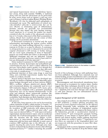
cosaminoglycans may be used to reduce adhesions and Figure 4.168. Gravitational device for the isolation of platelet‐
promote extracellular matrix quality, even though they rich plasma and platelet‐poor plasma.
have lower levels of evidence for success; they also carry
a low risk of complications. 40,49–51,54,59,60,67,71,80,82 Intra‐ or
perilesional injection of these same drugs is used less benefit of this technique in horses with pathology have
often, and controlled experimental studies have been not been published. Although these materials may aid
unable to confirm a benefit. tendon healing, proper rehabilitation and a convalescent
Several medical therapies have been developed spe- exercise program remain paramount to success with any
cifically for tendon or ligament injuries. Injectable autol- treatment.
ogous biologic therapies such as bone marrow, BMAC, Electromagnetic and electroshock stimulations have
MSCs, acellular collagen, platelet‐rich plasma (PRP), been proposed as treatments for tendon injury. Studies
and concentrated plasma are commonly used to treat suggest that these techniques should be used with cau-
SDF tendonitis (Figures 4.168 and 4.169). Acute, suba- tion because they can cause physical damage to the tis-
15
cute, or chronic tendinitis with a persistent centralized sue. Similarly, low‐level laser therapy (also called soft
4
hypoechoic area can be injected intralesionally. In the or cold laser therapy) has not substantiated clinical
acute and subacute phase, patient‐side products like claims of benefit.
PRP, BMAC, and allogeneic MSCs are used due to their
availability. Such therapies are likely beneficial even dur-
ing the acute phase as they help modulate the inflamma- Surgical Management of SDF Tendinitis
tory process. Two surgical procedures are described for treatment
At this time, bone marrow or fat can be harvested for of SDF tendinitis: a tendon splitting procedure and
culture expansion of MSCs, which requires 2–4 weeks. 71,124 proximal accessory ligament transection on the affected
Several experimental and clinical studies have described forelimb (frequently both, including the unaffected
the treatment of SDF tendonitis with adipose‐derived limb). The surgical technique of longitudinal tendon
stem cells in PRP, adipose‐derived stem cells, and splitting was advocated to decompress the lesion (relieve
91
30
bone marrow‐derived MSCs, 51,127 with improved tendon the hematoma), provide a vascularization channel to
organization and decreased re‐injury rates in racehorses central lesions, 2,140 and consequently promote healing,
reported. Use of ultrasound guidance for injection facili- particularly of core lesions in injured SDFT. In experi-
tates the injection into the lesion (Figure 4.169). mental and clinical studies, tendon splitting decreased
Intravenous or intra‐arterial regional limb perfusion the core lesion size within 8 weeks. 2,60 Importantly, ten-
using stem cells has been used clinically in some cases of don fibers are separated in the longitudinal plane and
extensive SDF tendinitis due to the difficulty of ade- not transected, with this procedure, or more fiber dam-
quately injecting the entire lesion. Although some stud- age could occur.
ies have evaluated the localization of stem cells following Transection of the accessory ligament (superior check)
regional perfusion, 128,131 studies evaluating the possible of the SDFT has been reported to drop the fetlock angle