
Page 621 - Adams and Stashak's Lameness in Horses, 7th Edition
P. 621
Lameness of the Distal Limb 587
swelling is paramount to limiting the fibrosis and ten- histologic lesions. If an SDFT injury is acute/subacute
don disfiguration that can be permanent. and has clinical signs of inflammation, a repeat ultra-
VetBooks.ir firm swelling on the palmar or plantar (flexor) surface tion has subsided. Hypoechoic areas within the SDFT
Chronic SDF tendinitis is manifested by fibrosis and
sound within 2 weeks is indicated after the inflamma-
can be edema fluid or hemorrhage early on after the
of the limb (Figure 4.164B). Chronic SDF tendinitis may
not have heat or edema associated with the condition, injury and may not necessarily be fiber disruption or
yet lameness may be present. The use of diagnostic ultra- structural damage (Figure 4.166). If the acute signs of
sonography enables initial and reasonably accurate injury have resolved and hypoechoic areas within the
determination of the extent and location of the lesions. SDFT persist, they likely represent fiber disruption
A complete ultrasound examination extends from (Figure 4.167). MRI has demonstrated an advantage
proximal to metacarpus/metatarsus to the heel bulbs over ultrasound for distinguishing fibrosis from normal
and determines the CSA of the SDFT and the lesions at tendon in chronic tendinitis. 67
multiple locations from proximal to distal. Comparison
to the contralateral side may provide a comparison and Treatment
possibly identify a subclinical condition. Longitudinal
scans permit the validation of structural lesions and the Immediate medical treatment of SDF tendinitis is rec-
assessment of fiber alignment. Hypoechogenicity can be ommended to reduce or eliminate the acute swelling and
caused by fibrillar disruption and/or inflammatory pro- inflammation as rapidly as possible to prevent fiber
cesses (hemorrhage, edema, and cellular infiltration in damage or early fibrous tissue formation. Routine phys-
the early stages). 107,116 Ultrasound images correlate to ical therapy such as icing, cryotherapy, pressure wraps,
Figure 4.166. Ultrasound longitudinal sections through the midportion of a normal SDFT (left) and SDFT with acute tendinitis. Note the
hypoechoic area (black) in the middle of the SDFT that usually corresponds to edema or hemorrhage in acute cases.
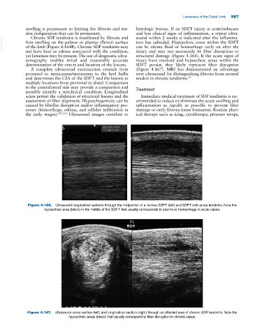
Figure 4.167. Ultrasound cross section (left) and longitudinal section (right) through an affected area of chronic SDF tendinitis. Note the
hypoechoic areas (black) that usually correspond to fiber disruption in chronic cases.