
Page 616 - Adams and Stashak's Lameness in Horses, 7th Edition
P. 616
582 Chapter 4
tendon (DDFT) in some cases. 36–39,41 Additionally, firm flexion exacerbates the lameness in 85% of horses with
digital pressure overlying the proximal SL in these cases hindlimb suspensory pain. 39
VetBooks.ir (Figure 4.161). The lameness is generally mild to moder- of the SL or avulsion fractures most frequently present
Horses with tearing of the Sharpey fibers at the origin
usually elicits a nonfatiguable painful response
with a history of acute onset of moderate to severe lame-
ate (1 to 2+ out of 5) at a trot and may be more obvious
when the horse is circled at a trot with the affected limb ness. Horses sustaining a fracture of the origin of the SL
on the outside of the circle. Horses with hindlimb PSD often have attained racing speeds in their workouts.
can exhibit moderate to severe hindlimb lameness. Digital pressure at the origin of the suspensory may
Lower limb flexion exacerbates the lameness in 50% of induce a painful response and exacerbate the lameness
horses with forelimb suspensory problems, and hock (Figure 4.161).
Horses with injury to the body or branches of the SL
usually have visible and palpable swelling and signs of
acute inflammation at the site of injury. In more insidi-
ous onset cases, the enlarged ligament has less inflam-
mation and is firmer on palpation. Pain on digital
pressure and a positive response to fetlock flexion are
common in affected horses.
Diagnostic Analgesia
Proximal suspensory injury can be challenging to
accurately diagnosis and requires a combination of
diagnostic analgesia and diagnostic imaging. Diagnostic
ultrasonography, nuclear scintigraphy, CT, and MRI can
all be used following lameness evaluation and diagnos-
tic analgesia to confirm and characterize SL pathology
(Figure 4.162). 22,41,44,72,143
Diagnostic analgesia is necessary to localize the lame-
ness to the proximal suspensory. There are many differ-
ent approaches to diagnostic analgesia of the proximal
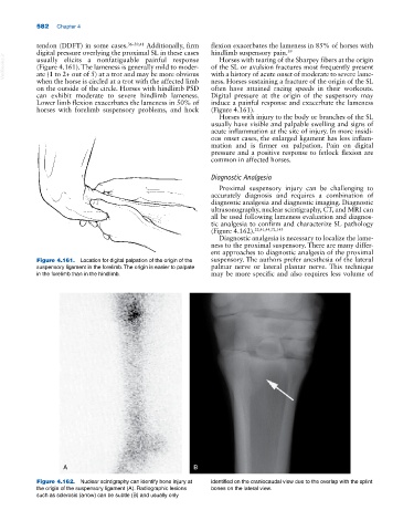
Figure 4.161. Location for digital palpation of the origin of the suspensory. The authors prefer anesthesia of the lateral
suspensory ligament in the forelimb. The origin is easier to palpate palmar nerve or lateral plantar nerve. This technique
in the forelimb than in the hindlimb. may be more specific and also requires less volume of
A B
Figure 4.162. Nuclear scintigraphy can identify bone injury at identified on the craniocaudal view due to the overlap with the splint
the origin of the suspensory ligament (A). Radiographic lesions bones on the lateral view.
such as sclerosis (arrow) can be subtle (B) and usually only