
Page 612 - Adams and Stashak's Lameness in Horses, 7th Edition
P. 612
578 Chapter 4
desmitis is present. This helps to define the contribution warranted. In conclusion, surgical removal of these dis-
of the fractured splint to the lameness. This is particu- tal splint fractures is often not necessary, but may be
VetBooks.ir on deep palpation of the bone, but the horse is quite 70% of horses with distal splint fractures have suspen-
performed, particularly for persistent fractures. At least
larly important in cases in which minimal pain is elicited
sory desmitis, which is generally agreed to be the cause
lame.
Radiographs should be taken in all cases to identify of continued lameness. 136
the fracture, its limits, and whether sequestration and
osteomyelitis exist in association with a complicated Closed Nonarticular and Nondistracted Proximal
fracture. The proximal fractures may extend toward or
into the carpometacarpal or tarsometatarsal joint Comminuted Fractures
(Figure 4.155). Closed nonarticular and nondistracted proximal com-
minuted fractures of the splint bones may heal success-
fully with 2–4 months of rest. In hindlimb lateral
Treatment
metatarsal fractures, the horses will be minimally lame
Small Distal Fractures and training can continue after the acute inflammation is
resolved (approximately 30 days). Surgery may become
Small distal fractures of the splint bones are tradi- necessary if:
tionally treated by removing the distal fragment, but this
approach is not universally recommended. 35,136 Up to 1. Draining tracts develop.
two‐thirds of the length of the splint can be removed 2. Lameness and pain associated with the fracture are
without untoward sequelae; however, distal splint frac- moderate to marked.
tures can heal spontaneously and are not usually a con- 3. Exuberant bony callus impinges on the SL.
tinued cause of lameness. Nonunion distal splint 4. Infection is present.
fractures are frequently not the cause of lameness, and 5. A nonunion or sequestra is developing on follow‐up
evaluation of the concomitant suspensory desmitis is radiographs.
6. A faster recovery and return to performance is desired.
If surgery is required, it should be performed aseptically
with the horse placed in lateral or dorsal recumbency.
Two surgical approaches have been successful:
removal of the fractured fragments only or removal of
the fracture and distal splint bone. If the fracture is in
the proximal third, any contiguous piece of bone present
should remain to stabilize the proximal component. In
closed fractures in which more than two‐thirds of the
splint bone is to be removed, a small bone plate is rec-
ommended to stabilize the remaining proximal frag-
ment. 17,101 If the proximal fragment is not anchored,
excessive movement of this fragment may occur and
result in interosseous desmitis, degenerative OA of the
carpometacarpal or tarsometatarsal joints, or avulsion
of the proximal fragment. Screw fixation is less fre-
quently successful. The small metacarpal bones are more
predisposed to avulsion after proximal fractures than
the metatarsal bones because of the major attachments
of the collateral ligaments.
Open Distal and Middle Fractures
Open distal and middle fractures of the splint bone
often lead to draining tracts or sequestra, so it is recom-
mended to treat these surgically with open removal of
the fracture with or without the distal splint
(Figure 4.156). The distal splint bone is left if it seems to
provide some stability to the proximal fragment.
Open Proximal Fractures
Open proximal fractures of the splint bones can be
more difficult to treat due to draining tracts, sequestra,
septic arthritis, and unstable proximal fragments.
Several treatment options are available including medi-
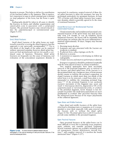
Figure 4.155. A dorsomedial to palmarolateral oblique cal management, fracture debridement, internal fixa-
101
radiograph of the carpus showing an oblique articular fracture of the tion, and complete removal if it involves the fourth
10
second metacarpal bone. metatarsal bone (Figure 4.157).