
Page 611 - Adams and Stashak's Lameness in Horses, 7th Edition
P. 611
Lameness of the Distal Limb 577
Etiology
Fractures of the distal part of the small metacarpal or
VetBooks.ir metatarsal bone result from external and internal trauma.
External trauma can result from a kick from another
horse, interference, direct blows from hitting another
object, or traumatic wounds. Internal trauma occurs from
increased axial compression forces on these bones during
races or from pressure from the SL or increased tension
from the fascial attachments. It is conjectured that the
increased incidence of left fourth metacarpal bone and
right second metacarpal bone fractures observed in
Thoroughbreds may be the result of increased weight‐
bearing on the bones when they are racing in a counter-
clockwise direction. In contrast, the SL and supporting
fascia may put these bones under tension sufficient to
cause fracture in the hindlimbs. 3,17,31,32,35,136 Because there
is an increase in incidence of left second metatarsal bone
and right fourth metatarsal bone, which is the tension
side of the hindlimb in horses that run counterclockwise,
it is logical to assume that tension created by the bow-
string effect of the SL or increased tension developed by
the internal fascia may lead to fracture.
It is difficult to decide whether the incidence of SL
desmitis is a result of distal splint bone fractures that
may cause irritation to this structure, or, conversely, that
the swollen SL becomes space occupying enough to
cause these fractures. Whatever the case, a higher inci-
dence of SL desmitis is noted in the forelimb in associa-
tion with distal splint bone fractures. 3,17,31,32,35,136
More complicated fractures of the proximal part of
the small metacarpal or metatarsal bones result from
direct trauma, either from interference or direct blows
to the surface. These fractures are often open initially,
which frequently results in osteomyelitis or septic arthri-
tis of the carpometacarpal or tarsometatarsal joint. In
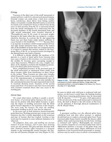
some cases there is not a break in the skin initially, but Figure 4.154. This lateral radiograph was taken 3 months after
the comminuted fractures become sequestered and result occurrence of a fourth metatarsal fracture. A large callus had
in recurrent draining tracts. Fractures of the second developed and there is minimal bridging of the fracture line. Source:
metacarpal bone of racehorses also have been associated Courtesy of Dr. Gary Baxter.
with excessive torsional forces that may occur in the
starting gate.
In cases in which only mild pain is evidenced with pal-
Clinical Signs pation, yet the horse is quite lame, the limiting features
of this horse resuming performance may be associated
On visual observation, swelling is usually a promi- with SL desmitis. 3,17,31,32,35,136 To gain a full appreciation
nent feature of proximal splint fractures, but it may or of the involvement of the small metacarpal or metatarsal
may not be present with distal splint fractures. In gen- bone, the limb is flexed so the full extent of this bone
eral, the degree of swelling associated with the distal can be palpated. A thorough physical examination of
splint fracture depends on the acuteness of the fracture. the SL should follow.
The more acute the fracture, the more the swelling.
Associated swelling in the SL may also be observed. Diagnosis
In the acute case, in both instances, horses frequently
point their foot. Trotting exercises may or may not cause A persistent swelling over the affected splint bone,
lameness, but this is totally dependent on the acuteness exhibiting heat and pain when pressure is applied,
and type of fracture that has resulted. Circling or fast should lead one to suspect a fractured splint bone. Some
work may be required to cause sufficient lameness to be fractured splint bones closely resemble the disease called
observed. splints. Some such fractures heal, but the bony swelling
On palpation, heat, pain, and swelling are obvious is confused with “splints.” Radiographs are necessary
features of acute fractures, and in some cases draining for a positive diagnosis of a fractured splint bone and to
tracts are also present. The pain and heat decrease with differentiate between a fracture of the splint bone and
time. However, because callus formation is a frequent splints or soft tissue injury. Direct infiltration of local
sequela to the nonsurgically treated fracture, the frac- anesthesia of the fractured small metacarpal or metatar-
ture site will become enlarged over time (Figure 4.154). sal bone may be used in cases in which a concomitant SL