
Page 608 - Adams and Stashak's Lameness in Horses, 7th Edition
P. 608
574 Chapter 4
VetBooks.ir
A B
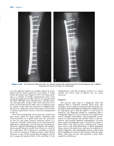
Figure 4.149. This transverse metatarsal fracture in a foal was repaired with a single broad DCP (A) and healed well with a relatively
large callus (B). Source: Courtesy of Dr. Gary Baxter.
over the affected region are usually present in active inflammation, and this normally resolves to a much
splints. Splints most commonly occur about 3 inches smaller size. Some cases of splints may not cause
below the carpal joint (Figure 4.150). One large swell- lameness.
ing or a number of smaller enlargements may occur
along the length of the splint bone at its junction with
the third MC/MT bone. Horses have variable lame- Diagnosis
ness but generally worsen with work and seem to be The obvious signs lead to a diagnosis when the
worse on hard ground. In mild cases no lameness may affected limb is examined carefully. Heat, pain, and
be evident at the walk, but lameness is exhibited dur- swelling over the regions mentioned, plus lameness, are
ing the trot. Palpation of the axial aspect of the splint enough to make the diagnosis. Diagnostic analgesia may
is useful to determine if there is obvious impingement be required to prove that the splint is a true cause of
of the SL. lameness. The diagnosis should be confirmed with a
New bone growth that occurs near the carpal joint radiograph (Figure 4.151) as fractures of the splint bone
may cause carpal OA (knee splints). Extensive new can be confused with splints. Ultrasonographic exami-
bone formation on a splint bone may also encroach nation can demonstrate concomitant injury to the sus-
on the SL and cause chronic lameness unless it is pensory and possible ligament impingement. In some
removed. Growths of this kind can be determined by cases, nuclear scintigraphy may be needed to confirm a
palpation, radiographs, and ultrasound examination. blind splint. New bone growth resulting from trauma
After the original inflammation subsides, the enlarge- may occur on the third metacarpal or third metatarsal
ments usually become smaller but firmer as a result of bones, close to the splint bone, and may be mistaken for
the ossification. The reduction in swelling is usually splints. Palpation and radiographs, however, show that
the result of resolution of fibrous tissue, rather than a these swellings are dorsal to the junction with the splint
decrease in size of the actual bone formation. In the bones. This type of new bone growth is most often
early stages, the greatest bulk of the swelling is from caused by interference.