
Page 604 - Adams and Stashak's Lameness in Horses, 7th Edition
P. 604
570 Chapter 4
VetBooks.ir
A B
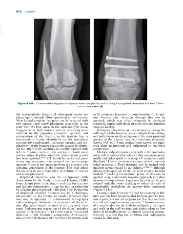
Figure 4.146. Craniocaudal radiograph of a displaced lateral condylar fracture in a racing Thoroughbred (A) repaired with three 5.5‐mm
cortical bone screws (B).
the supracondylar fossa, and subsequent screws are as P1 eminence fractures or comminution of the pal-
placed approximately 20 mm proximal to the first one. mar fracture line. Articular damage also can be
Most lateral condylar fractures can be repaired with assessed, which may affect prognosis in displaced
two screws. Ideal screw placement is parallel to the fractures, particularly those of more chronic duration
joint with the first screw in the supracondylar fossa, (days to weeks).
engagement of both cortices without depositing bone In displaced fractures, an open incision extending the
material in the opposing collateral ligament, and full length of the fracture, use of multiple bone clamps,
compression of the fracture so the fracture line is and initial focus on the reduction of the most proximal
eliminated or barely identifiable on the immediate portion of the fracture may help maximize reduction.
postoperative radiograph. Increased duration and dis- Screws (4.5‐ or 5.5‐mm cortical bone screws) are tight-
placement of the fracture reduce the success in obtain- ened distal to proximal and retightened to maximize
ing the latter result. Fractures are usually repaired with compression.
4.5‐ or 5.5‐mm cortical bone screws, although some Medial condylar fractures, especially in the hindlimbs,
success using headless titanium compression screws are at risk of catastrophic failure if they propagate prox-
has been reported. 47,105 CT should be performed prior imally and either spiral or develop a Y component mid‐
to starting the surgery to understand the fracture prop- diaphysis. Lateral condylar fractures can uncommonly
3
agation if there is any question about the presence of a spiral proximally. These fractures can be treated with
spiraling component to the fracture. This may affect multiple screws placed in lag fashion, 119,126,144 although
the decision to use a bone plate in addition to screws plating techniques are likely the most reliable fixation
and screw placement. method. Locking compression plates (LCPs) can be
53
Displaced fractures can be compressed with applied using a minimally invasive approach to reduce
Association for the Study of Internal Fixation (ASIF) incisional complications. Lag screw fixation can be per-
bone clamps prior to drilling to stabilize the fracture formed with the horse standing to reduce the risk of
and initiate compression or can be held in reduction catastrophic breakdown on recovery from anesthesia
by a Steinmann pin placed in the glide hole. Reduction (Figure 4.145).
of displaced condylar fractures can be a challenge, Casting is usually not performed for recovery. A full‐
particularly in the cranial to caudal direction that limb cast has been recommended for long spiraled frac-
may not be apparent on craniocaudal radiographs ture repairs, but not all surgeons use this because there
taken at surgery. Arthroscopic evaluation of the artic- is a risk of complications in recovery. Horses are usu-
110
ular alignment should be used in all displaced frac- ally comfortable on the limb immediately after repair
tures. Arthroscopic inspection of the dorsal articular and walk in the stall without gait deficit when on low
surface often reveals a 1‐ to 2‐mm step in the cranial doses of phenylbutazone. Continued lameness postop-
direction of the fractured component. Arthroscopy eratively is a red flag for problems and radiographs
also allows debridement of other bone fragments such should be obtained.