
Page 725 - Adams and Stashak's Lameness in Horses, 7th Edition
P. 725
Lameness of the Proximal Limb 691
antimicrobials. Chronic cases of TS sepsis are candi- LDFT Tendinitis
9
dates for tenoscopic evaluation. Surgical intervention in Primary injury to the LDFT is a rarely reported cause
VetBooks.ir drainage of fluid, placement of indwelling drains, TS of hindlimb lameness in performance horses but has
such cases may involve incision into the sheath to allow
recently been reported in 4 horses that had MRI exami-
tenoscopy, and transection of the tarsal flexor retinacu-
32
lum. Bony abnormalities involving the ST or tuber cal- nation of the tarsal region (Figure 5.93). Lameness was
mild/moderate in severity and insidious in onset in all
canei should be treated as the primary source of horses. Responses to flexion tests were variable. TS effu-
infection and addressed surgically. The prognosis for sion was slight in three horses and mild/moderate in one
return to athletic function is frequently guarded and horse. Two horses were positive to DBLPN block, but all
often requires a protracted course of treatment. Further horses showed improvement (70%–90%) in lameness
information on treating synovial sepsis can be found in after TS analgesia (US‐guided injection). Radiographic,
Chapter 12. scintigraphic, and ultrasonographic findings were
A B
Figure 5.92. (A) Oblique radiographs of two different horses demonstrating an acute, open (A) and chronic, closed (B) fracture of the
sustentaculum tali (arrows).
A B
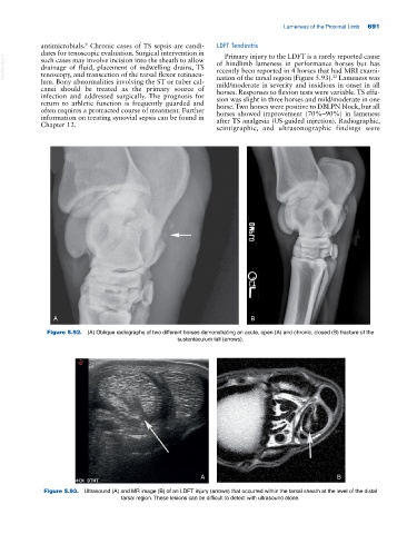
Figure 5.93. Ultrasound (A) and MR image (B) of an LDFT injury (arrows) that occurred within the tarsal sheath at the level of the distal
tarsal region. These lesions can be difficult to detect with ultrasound alone.