
Page 727 - Adams and Stashak's Lameness in Horses, 7th Edition
P. 727
Lameness of the Proximal Limb 693
VetBooks.ir
A B
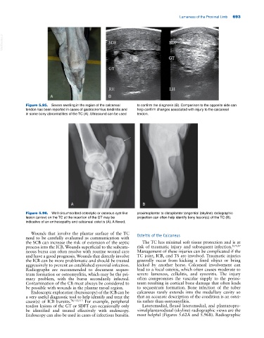
Figure 5.95. Severe swelling in the region of the calcaneal to confirm the diagnosis (B). Comparison to the opposite side can
tendon has been reported in cases of gastrocnemius tendinitis and help confirm changes associated with injury to the calcaneal
in some bony abnormalities of the TC (A). Ultrasound can be used tendon.
A B
Figure 5.96. Well‐circumscribed osteolytic or osseous cyst‐like proximoplantar to distoplantar tangential (skyline) radiographic
lesion (arrow) on the TC at the insertion of the GT may be projection can often help identify bony lesion(s) of the TC (B).
indicative of an enthesopathy and calcaneal osteitis (A). A flexed,
Wounds that involve the plantar surface of the TC Osteitis of the Calcaneus
need to be carefully evaluated as communication with
the SCB can increase the risk of extension of the septic The TC has minimal soft tissue protection and is at
process into the ICB. Wounds superficial to the subcuta- risk of traumatic injury and subsequent infection. 8,70,87
neous bursa can often resolve with routine wound care Management of these injuries can be complicated if the
and have a good prognosis. Wounds that directly involve TC joint, ICB, and TS are involved. Traumatic injuries
the ICB can be more problematic and should be treated generally occur from kicking a fixed object or being
aggressively to prevent an established synovial infection. kicked by another horse. Calcaneal involvement can
Radiographs are recommended to document seques- lead to a focal osteitis, which often causes moderate to
trum formation or osteomyelitis, which may be the pri- severe lameness, cellulitis, and synovitis. The injury
mary problem, with the bursa secondarily infected. often compromises the vascular supply to the perios-
Contamination of the CB must always be considered to teum resulting in cortical bone damage that often leads
be possible with wounds in the plantar tarsal region. to sequestrum formation. Bone infection of the tuber
Endoscopic exploration (bursoscopy) of the ICB can be calcaneus rarely extends into the medullary cavity so
a very useful diagnostic tool to help identify and treat the that an accurate description of the condition is an ostei-
cause(s) of ICB bursitis. 74,112,113 For example, peripheral tis rather than osteomyelitis.
tendon lesions of the GT or SDFT can occasionally only Lateromedial, flexed lateromedial, and plantaropro-
be identified and treated effectively with endoscopy. ximalplantarodistal (skyline) radiographic views are the
Endoscopy can also be used in cases of infectious bursitis. most helpful (Figures 5.62A and 5.96B). Radiographic