
Page 729 - Adams and Stashak's Lameness in Horses, 7th Edition
P. 729
Lameness of the Proximal Limb 695
endoscopy of the bursa allows surgical debridement of of the bursa dorsal to the insertion of the GT that may
the torn tissue and exploration of the calcaneal bursa for communicate with the ICB. Ultrasonography may be
VetBooks.ir the tendon also have disruption of the tendon fibrocarti- defined margins, focal or diffuse anechoic to hypoechoic
used to best detect enlargement of the tendon, poorly
any other damage. Horses with unstable displacement of
lesions, and loss of normal fiber alignment (diffuse >
lage cap. Removal of this tissue (torn retinaculum and
the damaged fibrocartilage cap) results in stable subluxa- focal). The lesions seem to occur predominantly in the
tion and can return horses to athletic activity. Both lesions body of the tendon and less frequently at the insertion
can be detected with preoperative ultrasonography. 149 on the TC. In the majority of horses, however, GT occurs
With conservative management the prognosis for without concurrent tendinous or ligamentous injury.
breeding soundness or light pleasure riding is usually Severe injuries may allow the point of the hock to “drop”
good for stable luxations. Realistically, too few cases because the reciprocal apparatus is no longer func-
have been treated surgically with long‐term follow‐up to tional. 76,85,142 Radiographs should be obtained for the
make an objective comment about the prognosis for ath- detection of a fracture of the calcaneus that can manifest
letic ability, because every case is different. a similar appearance. Diagnostic US can help confirm
the location of the injury but is not necessary for an
accurate diagnosis. Conservative treatment with stall
Gastrocnemius Tendinitis
rest and controlled exercise for 6–12 month is usually
The SDFT begins medial to the GT and rotates/ indicated. 85,142 Rehabilitation protocols can include
courses from medial to caudal to attach to the calcaneus water treadmill exercise. Horses with mild to moderate
before continuing distally. The LDFT is deep to both the lesions have a reasonable prognosis for return to athletic
SDFT and GT and courses distally over the plantarome- work, but the prognosis for horses with more severe
dial aspect of the calcaneus and the ST. The combined lesions is guarded.
SDFT and GT form the common calcaneal tendon, Horses with chronic calcaneal bursitis often have
which is the major extensor of the tarsus. Severe injuries insertional changes of the GT. Chronic calcaneal tend-
to the common calcaneal tendon can cause partial or initis may have osteolytic lesions at the insertion of the
complete loss of support to the tarsus. Lameness and GT on the TC (“gastrocnemius enthesitis”). Well‐cir-
swelling in the region of the distal common calcaneal cumscribed osteolytic or osseous cyst‐like lesion(s) on
tendon are most commonly due to tendonitis of the GT, the TC at the insertion of the GT may be associated with
which is considered a rare cause of hindlimb lameness in enthesopathy or a calcaneal osteitis as a component of
the horse. 41,114 Lameness may be sudden or gradual in focal nonseptic osteitis. A radiographic examination of
onset, and the severity of lameness varies depending on the tarsus should include a flexed, proximoplantar to
the injury. Lameness is usually exacerbated by a proxi- distoplantar tangential (skyline) view, and a flexed lat-
mal limb flexion test. Lameness is usually improved by eral radiographic view of the calcaneus is advantageous
perineural analgesia of the tibial nerve. for identifying this lesion and is recommended for all
In association with enlargement of the GT, there can horses with calcaneal bursitis. Radiographic signs
be a mild to moderate enlargement of the calcaneal include increased soft tissue swelling, osseous lysis, frag-
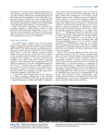
bursa (ICB and CB) (Figure 5.98). This can give the limb ments/sequestra, and new bone production more com-
a “capped” appearance possibly attributable to distension monly associated with chronic conditions.
A B
Figure 5.98. Thickening and swelling was identified in the hypoechoic areas with loss of normal fiber alignment on both the
calcaneal tendon of this horse (A). Ultrasound demonstrated transverse and longitudinal planes (B).
enlargement of the gastrocnemius tendon with focal and diffuse