
Page 760 - Adams and Stashak's Lameness in Horses, 7th Edition
P. 760
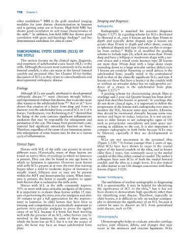
726   Chapter 5


            other modalities.  MRI is the gold standard imaging   Imaging and Diagnosis
                           38
            modality for joint disease characterization in humans   Radiography
  VetBooks.ir  shown good correlation to soft tissue characteristics of   (Figure 5.127). In a grading scheme for SCLs developed
            and is gaining some use in horses. High‐field MRI has
                                                                  Radiography is standard for accurate diagnosis
            the stifle.  In addition, low‐field MRI has shown good
                    11
                                                   50
            correlation with gross and histologic findings  and has   by Howard et al., type 1 lesions are less than 10 mm in
            demonstrated usefulness for clinical cases. 35     depth and typically dome shaped; type 2 lesions are
                                                               greater than 10 mm in depth and either dome, conical,
                                                               or spherical shaped; and type 3 lesions are flat or irregu-
            SUBCHONDRAL CYSTIC LESIONS (SCLS) OF               lar bone surface.   Wallis et  al. modified the grading
                                                                               25
            THE STIFLE                                         scheme to include type 2A, which are more than 10 mm
                                                               deep and have a lollipop or mushroom shape with a nar-
              This section focuses on the clinical signs, diagnosis,   row cloaca and a round cystic lucency; type 2B lesions
            and treatment of subchondral cystic lesion (SCL) in the   are more than 10 mm deep with a large dome shape
            stifle. Although this occurs most commonly in the medial   extending down to a large articular surface defect; type
            femoral condyle, it also can occur in the lateral femoral   3 lesions are condylar flattening or small defects in the
            condyle and proximal tibia. See Chapter 10 for further   subchondral bone, usually noted in the contralateral
            discussion of SCLs as they relate to osteochondrosis and   limb to that of the clinically significant SCL; and type 4
            developmental orthopedic disease.                  lesions are those that have a lucency in the condyle with
                                                               or without an articular defect but no radiographic evi-
            Etiology                                           dence of a cloaca in the subchondral bone plate
                                                               (Figure 5.127). 60
              Although SCLs are usually attributed to developmental   A grading scheme for characterizing presale films in
            orthopedic disease, 28,31  some clinicians strongly believe,   yearlings and 2‐year‐old Quarter horses has been devel-
            and experimental evidence has shown, that it can also occur   oped that describes lesions in general.  In yearlings that
                                                                                                5
            after trauma to the subchondral bone. 44,59  Ray et al.  have   do not show clinical signs, it is important to follow the
                                                      44
            shown that creation of a lesion 3 mm deep and 5 mm in   progression of the lesions with radiographs over time to
            diameter into the subchondral bone created SCL in 5 of 6   monitor the SCL, because some may resolve. Likewise,
            horses. Furthermore, von Rechenberg et al.  showed that   they should be monitored clinically because some may
                                                45
            the lining of the cysts contains significant inflammatory   worsen and begin to induce lameness. It is not uncom-
            mediators that may be responsible for enlargement and   mon  in  older  horses  to  see  radiographic  signs  of  OA
            persistence of the cyst. The tissues from postmortem cystic   such as periarticular osteophytes and joint space nar-
            material had significant osteoclastic function on bone.   rowing in addition to the cyst. It is also important to
            Therefore, regardless of the cause of cyst formation, persis-  compare radiographs in both limbs because SCLs may
            tent enlargement of some lesions may be due to a vicious   be bilateral, especially  if they are developmental  in
            cycle of inflammation.                             origin.
                                                                  SCLs are  not uncommon in  the proximal tibia
                                                                            56
            Clinical Signs                                     (Figure 5.128).  In horses younger than 2 years of age,
                                                               tibial SCLs have been shown to occur in the cranial
              Horses with SCL of the stifle can present in several   aspect of the lateral condyle of the tibia, and in horses
            different ways. Historically, many of these lesions are   older than 2 years, they commonly occur in the medial
            found on survey films of yearlings in which no lameness   condyle of the tibia, either cranial or caudal. The author’s
            is present. They can also be found in any age horse in   colleagues have seen SCLs of both the medial femoral
            which no lameness is apparent. However, most horses   condyle and the tibia in a single horse. It is also typical
            with stifle SCLs present at or near the time that training   in older horses to see OA changes concurrent with these
            begins when a mild to moderate degree of lameness is   lesions (Figure 5.128).
            usually found. Effusion may or may not be present
            within the MFT and femoropatellar joints. When lame-  Nuclear Scintigraphy
            ness is present, the horse is usually positive to upper
            limb flexion that is isolated to the stifle joint.    The usefulness of nuclear scintigraphy in diagnosing
              Horses with SCL in the stifle commonly improve   SCL is questionable. It may be helpful for identifying
            50% or more with intra‐articular analgesia of the joints.   the significance of SCL in the tibia,  but it has not
                                                                                                56
            It is important to evaluate these horses within 15 min-  been shown to demonstrate high specificity other than
            utes of performing the intra‐articular analgesia and after   for identifying joint disease.  Therefore, especially in
            30 minutes to get a full appreciation for the improve-  older horses, it is difficult to rely on nuclear scintigra-
            ment in lameness. In older horses that have been in   phy to determine the significance of an SCL because it
            training and competition, it is particularly important to   cannot be used to discern an SCL from generalized
            perform intra‐articular analgesia to prove the signifi-  joint disease, such as OA.
            cance of an SCL because if a horse has been working
            well with the presence of an SCL, other factors may be   Ultrasonography
            involved in the lameness. In some of these cases, in
            which the horse has an SCL but it is not the source of   Ultrasonography helps to evaluate articular cartilage
            pain, the horse may have an intact subchondral bone   surface, joint  effusion, debris,  and changes that may
            plate.                                             occur in the meniscus and cruciate ligaments. SCLs