
Page 765 - Adams and Stashak's Lameness in Horses, 7th Edition
P. 765
Lameness of the Proximal Limb 731
VetBooks.ir
A
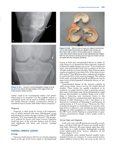
Figure 5.132. Menisci from a 5‐year‐old cutting horse that had
chronic left medial femorotibial pain (Bottom) and a 9‐year‐old
Warmblood with no history of femorotibial pain for comparison
(Top). Both bottom menisci appear to lack substantial axial integrity,
with the right meniscus showing axial damage and striation (arrow)
compared with the normal top meniscus.
lesions in foals and osteochondral disease in adults. In
foals, Hance et al. showed that three separate categories
of femoral condyle lesions can occur. Type I lesions are
21
septic, and subchondral bone lysis is present in the face
of septic arthritis and osteomyelitis. Type II lesions show
osseous irregularity in less than 50% of the femoral con-
dyle surface. Type III lesions show widespread irregular-
ity and some have a thin osseous fragment. The authors
of the study concluded that type II and III lesions may be
some form of developmental orthopedic disease or vas-
B cular insult.
Osteochondral lesions of the femoral condyles in
adult horses nearly always involve the medial femoral
Figure 5.131. Caudal to cranial radiographic images of acute condyle. These lesions are usually considered to be
(A) and more chronic (B) fragmentation of the origin of the long traumatic in origin; however, there is growing concern
digital extensor tendon (arrows).
that some of the lesions may be developmental in origin
because abnormalities in condylar shape, such as dim-
surface needs to be reconstructed and/or vital attach- pling or flattening, may predispose horses to this
61
ments need to be retained. For the condylar surface, a problem. However, a postmortem study showed no cor-
cannulated screw can be used to stabilize a fracture of relation of MFC flattening to OA, and Barret et al.
13
the medial femoral condyle. Conservative therapy is showed no correlation of flattening to performance in
sometimes best in horses with Salter–Harris fractures. 20 cutting horses at 3–4 years of age. However, the effect
5
of meniscal integrity on development of articular carti-
Prognosis lage lesions is unknown, and considering how meniscal
damage can affect prognosis for various lesions within
Prognosis is often good for horses with fragmenta- the MFT joint and the findings in young horses on post-
tion in which minimal soft tissue debridement occurs mortem examinations (Figure 5.132), further work is
and minimal secondary damage is present. For MICET needed. 14
48
fractures, 76% were sound after removal. The progno-
48
sis is reduced for horses with larger fracture fragments Clinical Signs and Diagnosis
involving more of the articular surfaces and for those
with significant soft tissue and articular cartilage Foals with type I and III lesions are typically severely
damage. 48 lame, and those with type II lesions are usually moder-
ately lame. However, all cause clinical signs that obvi-
ously point to a stifle problem. Radiographs typically
FEMORAL CONDYLE LESIONS reveal subchondral bone lucency indicative of the
Etiology damage (Figure 5.133).
For nonseptic cases, medial femoral condyle osteo-
Femoral condyle lesions fall into two broad categories chondral lesions are common in young western perfor-
based on the age of the horse: septic or developmental mance athletes; however, they can occur in any type of