
Page 770 - Adams and Stashak's Lameness in Horses, 7th Edition
P. 770
736 Chapter 5
VetBooks.ir
A
Figure 5.137. A caudal to cranial radiographic image showing
osteophytes on the medial aspect of the tibia and the intercondylar
eminence, which are common in many types of diseases involving
the femorotibial joints.
B
Figure 5.139. Ultrasonographic image (A) of a severe meniscal
A tear that correlated well with the gross appearance (B). Source:
Courtesy of Dr. Laurie Goodrich.
ultrasonographic findings since most of the meniscus is
inaccessible at surgery, leading to potential false‐positive
and/or false‐negative findings.
Other diagnostic techniques include nuclear scintig-
raphy, which has poor sensitivity and modest specificity
for stifle disease in general; 4,18 MRI, which is useful but
limited in availability; contrast CT, which requires com-
munication of the tear with the synovial cavity; and
arthroscopy, which is an important diagnostic tool that
is readily available but unable to visualize much of the
weight‐bearing surface of the meniscus. Therefore, the
lack of findings of a meniscal lesion with arthroscopy
B does not necessarily rule out the absence of a lesion.
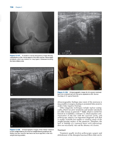
Figure 5.138. Ultrasonographic images of the medial meniscus Treatment
in both weight‐bearing (A) and non‐weight‐bearing positions (B).
Notice how the meniscal tear becomes more apparent in the non‐ Treatment usually involves arthroscopic surgery and
weight‐bearing position. debridement of the disrupted meniscal fibers that can be