
Page 774 - Adams and Stashak's Lameness in Horses, 7th Edition
P. 774
740 Chapter 5
FEMUR AND COXOFEMORAL REGION
VetBooks.ir niColas s. ernst and troy n. trumble
THE FEMUR Diagnosis
Conditions of the femoral region that contribute to Excessive movement of the distal limb is usually pre-
lameness are uncommon and can vary greatly in sent on palpation. Fractures of the distal growth plate
severity. For example, femoral fractures usually cause are usually evident because of displacement, instability,
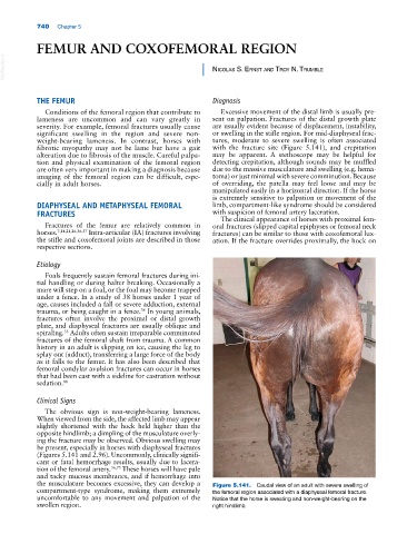
significant swelling in the region and severe non‐ or swelling in the stifle region. For mid‐diaphyseal frac-
weight‐bearing lameness. In contrast, horses with tures, moderate to severe swelling is often associated
fibrotic myopathy may not be lame but have a gait with the fracture site (Figure 5.141), and crepitation
alteration due to fibrosis of the muscle. Careful palpa- may be apparent. A stethoscope may be helpful for
tion and physical examination of the femoral region detecting crepitation, although sounds may be muffled
are often very important in making a diagnosis because due to the massive musculature and swelling (e.g. hema-
imaging of the femoral region can be difficult, espe- toma) or just minimal with severe comminution. Because
cially in adult horses. of overriding, the patella may feel loose and may be
manipulated easily in a horizontal direction. If the horse
is extremely sensitive to palpation or movement of the
DIAPHYSEAL AND METAPHYSEAL FEMORAL limb, compartment‐like syndrome should be considered
FRACTURES with suspicion of femoral artery laceration.
The clinical appearance of horses with proximal fem-
Fractures of the femur are relatively common in oral fractures (slipped capital epiphyses or femoral neck
horses. 7,14,21,26,36,57 Intra‐articular (IA) fractures involving fractures) can be similar to those with coxofemoral lux-
the stifle and coxofemoral joints are described in those ation. If the fracture overrides proximally, the hock on
respective sections.
Etiology
Foals frequently sustain femoral fractures during ini-
tial handling or during halter breaking. Occasionally a
mare will step on a foal, or the foal may become trapped
under a fence. In a study of 38 horses under 1 year of
age, causes included a fall or severe adduction, external
trauma, or being caught in a fence. In young animals,
36
fractures often involve the proximal or distal growth
plate, and diaphyseal fractures are usually oblique and
spiraling. Adults often sustain irreparable comminuted
35
fractures of the femoral shaft from trauma. A common
history in an adult is slipping on ice, causing the leg to
splay out (adduct), transferring a large force of the body
as it falls to the femur. It has also been described that
femoral condylar avulsion fractures can occur in horses
that had been cast with a sideline for castration without
sedation. 88
Clinical Signs
The obvious sign is non‐weight‐bearing lameness.
When viewed from the side, the affected limb may appear
slightly shortened with the hock held higher than the
opposite hindlimb; a dimpling of the musculature overly-
ing the fracture may be observed. Obvious swelling may
be present, especially in horses with diaphyseal fractures
(Figures 5.141 and 2.96). Uncommonly, clinically signifi-
cant or fatal hemorrhage results, usually due to lacera-
tion of the femoral artery. 36,73 These horses will have pale
and tacky mucous membranes, and if hemorrhage into
the musculature becomes excessive, they can develop a Figure 5.141. Caudal view of an adult with severe swelling of
compartment‐type syndrome, making them extremely the femoral region associated with a diaphyseal femoral fracture.
uncomfortable to any movement and palpation of the Notice that the horse is sweating and non‐weight‐bearing on the
swollen region. right hindlimb.