
Page 769 - Adams and Stashak's Lameness in Horses, 7th Edition
P. 769
Lameness of the Proximal Limb 735
Diagnosis meniscal injury can vary. In a study by Walmsley, 40%
of cases presented acutely, and 53% were insidious in
Radiographs are usually unremarkable unless the ori-
VetBooks.ir gin of the ligament avulses from the intercondylar fossa, 66% were positive to upper limb flexion, 47% had MFT
onset. The median lameness grade was 3 out of 10,
65
15
a midbody tear shows dystrophic mineralization, or a
joint and/or femoropatellar effusion, and 93% responded
medial tibial eminence fragment is present. However, the
65
latter finding is not pathognomonic for cranial cruciate to intra‐articular analgesia. However, meniscal injury
can be secondary to chronic OA or SCLs, and signs can
ligament damage because a fracture at the apex of the include capsule thickening of the MFT joint and disuse
eminence often does not involve the ligament. Ultrasound atrophy of the surrounding musculature.
gives variable results because the cranial cruciate liga-
ment area is difficult to image; however, a small section
of the cranial cruciate ligament insertion on the tibia can Diagnosis and Imaging
be visualized with the limb flexed. Contrast‐enhanced Radiographs can be normal in acute cases of primary
CT has some merit if the ligament tear communicates meniscal damage. However, damage to the meniscus
with the joint. MRI is available in limited locations and may lead to joint space narrowing, so it is important
38
probably demonstrates the best chances of acquiring an that these images be reassessed because poor positioning
accurate diagnosis. High‐field, short‐bore magnets can can lead to false interpretation of the radiographs. In
58
accommodate some horses for accurate imaging. Low‐ addition, radiographic abnormalities often accompany
field scanners are gaining favor and have shown promise meniscal tearing in more chronic cases, as has been
35
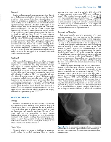
for accurate diagnosis. Arthroscopic surgery can be shown in several studies 4,12,62 Mineralization of the
used to explore the joint and assess the extent of damage meniscus (Figure 5.136) and osteophytes on the medial
and is currently the best method to accurately diagnosis tibial plateau (Figure 5.137) and intercondylar eminence
cranial cruciate damage. 39,42 of the tibia are common in horses with more chronic
lameness. In updated results from Walmsley, 23% of
64
Treatment horses with meniscal damage had new bone formation
at the medial intercondylar eminence of the tibia, and
Osteochondral fragments from the tibial eminence this was associated with cranial cruciate injury in only
can be removed and damaged tissues debrided arthro- 7% of cases. 34
scopically. Microfracture may help stimulate endoge- Ultrasonographic findings can include characteriza-
nous repair of avulsed ligament from bone in the tion of an abnormal size, thickness, variability in echo-
intercondylar space. Damaged tissue can be debrided, genicity, location of the meniscus, prolapse of the
and although no repair technique has been successfully meniscus medially, and overt meniscal tearing. 4,8,12,23 It is
developed for the horse, partial tears can be augmented important when assessing for a tear that the area is
with platelet‐rich plasma (PRP) or mesenchymal stem imaged in both weight‐bearing and non‐weight‐bearing
cells injected into the tissues or joint. Other therapies stances (Figures 5.138 and 5.139). Non‐weight‐bearing
39
to consider include intra‐articular stem cell or IRAP imaging often causes opening of the meniscal tear for
therapy to use the growth factor properties that these better demonstration of the defect. For acute severe inju-
two treatments are meant to induce. Overall, the prog- ries, the cranial cruciate ligament and medial collateral
nosis depends on the extent of damage, but is often con- ligament may also be involved, but it is not uncommon
sidered poor. However, some horses have returned to to see isolated meniscal injuries. Despite its widespread
work. 29,39,43 Caudal cruciate ligament injuries have been use to diagnose meniscal lesions, it is difficult to validate
reported, and most of them involve longitudinal fraying
and not true ligament rupture. 3,8,36,41,47
MENISCAL INJURIES
Etiology
Meniscal lesions can be acute or chronic. Acute dam-
age can occur with a bad step or an accident that leads
to shifting or shear forces between the femur and tibia.
Lesions are often seen in other structures such as cruci-
ate ligaments and articular cartilage. Horses of any
breed and use are susceptible to these lesions.
Degenerative lesions of meniscus are seen to associate
positively with articular cartilage degeneration and oste-
ophyte scores. Pathogenesis is unknown. Both lateral
14
and medial meniscal lesions occur, but damage to the
medial meniscus is most common. 14
Clinical Signs Figure 5.136. A lateromedial radiographic image demonstrating
mineralization of the caudal aspect of the meniscus. Arthroscopic
Meniscal injuries are acute or insidious in onset and removal of this mineralization is rarely indicated because the OA is
usually affect the medial meniscus. Signs of medial often advanced.