
Page 771 - Adams and Stashak's Lameness in Horses, 7th Edition
P. 771
Lameness of the Proximal Limb 737
seen. In addition, it is common to test the looseness of Clinical Signs and Diagnosis
the meniscus with a probe during surgery. The Horses with synovitis of the femorotibial joints may
VetBooks.ir seems to be associated in some cases with damage and not be lame, but have mild to moderate effusion in the
significance of this finding is unknown, but subjectively
MFT joint. These horses may show some response to
protrusion of the meniscus identified on ultrasound. In
addition, manipulation of the meniscus permits better flexion and a gait that can be described as stiff. Some
visualization of the tibia for evidence of secondary horses can compete well with this finding, but they
damage. should be monitored closely. As mentioned previously,
Walmsley developed a grading scale for meniscal all diagnostic techniques can be negative in these cases,
injuries: grade I is characterized by axial tearing through including arthroscopic surgery.
the cranial ligament of the medial meniscus and into the Horses with synovitis secondary to other primary
meniscus, grade II is the same as grade I but with torn lesions within the MFT joint usually have a history of a
tissue and visible extent of the damage, and grade III is a short response period to intra‐articular medication.
severe tear that extends beneath the femoral condyle. 65 However, trainers and owners usually note that the
In an updated review of horses with meniscal injuries, horse either never fully regained its level of performance
Walmsley found that of 126 cases, 111 were medial and or constant medication was needed. In these cases, addi-
25 were lateral and 53% were grade I, 28% grade II, tional diagnostics, including diagnostic arthroscopy, are
63
and 17% grade III. In that study, 76% had articular often required to fully characterize the problem.
cartilage damage, and 14% had damage to the cranial Horses with significant OA of the femorotibial joints
cruciate ligament. Of those with medial meniscal tear- are usually lame at the walk with loss of muscle mass
ing, 45% were sound compared with 75% of those with and soft tissue swelling and effusion of the MFT joint.
lateral meniscal involvement. Sixty percent of horses Concurrent abnormalities such as OA, SCL of the medial
with grade I tearing were sound compared with 65% of femoral condyle, and medial meniscal damage are com-
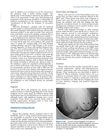
those with grade II and 10% of those with grade III. mon (Figure 5.140). Although a history of predisposing
Overall, 51% of the horses were sound with surgery. injury and disease of the stifle is usually noted, some
63
Similarly, Cohen et al. found decreasing prognosis with horses may develop this problem insidiously without a
increasing meniscal damage, with no horses with grade notable injury.
9
III tearing returning to previous use. Because most of
the meniscus cannot be visualized arthroscopically, a Treatment
tear or lesion in the body of the meniscus may be treated
by extracapsular injection of stem cells or PRP into the Horses with synovitis usually respond well to intra‐
lesion(s) or into the joint. Horses with meniscal lesions articular medication, topical anti‐inflammatories,
treated with bone marrow derived stem cells returned to systemic medication, and extracorporeal shockwave
work more often than those historical controls. If the therapy. These medications are described in depth in
16
tear is visible arthroscopically, then various suturing and Chapter 8. Horses typically respond well to rest or a
40
staple techniques are also available. In general, sutur- reduction in training, which are often recommended.
ing is preferred over stapling. The efficacies of these lat- However, failure to respond to medication and recur-
ter techniques to treat meniscal lesions are currently rence of lameness are often the key findings that lead
unknown. to more intensive imaging and possibly diagnostic
arthroscopy.
Prognosis
As noted above, the prognosis for lesions of the
medial meniscus is relatively guarded, but for the lateral
meniscus can be quite good. In addition, horses with
small lesions that can be debrided can do relatively well,
although horses with significant tearing carry a poor
prognosis. Great efforts are currently being made to
provide augmentative therapies, such as stem cells,
which have shown some promise. 16
SYNOVITIS/CAPSULITIS/OA
Etiology
Horses that undergo chronic repetitive stress to the
hindlimbs, such as young western performance horses,
are susceptible to synovitis in the femorotibial joints,
especially the medial compartment. This is similar to
racehorses that develop synovitis in their metacarpophalan- Figure 5.140. Caudal to cranial radiograph of the stifle of a
geal and carpal joints. A primary source of synovitis is 6‐year‐old Quarter horse mare with multiple radiographic abnormali-
usually not apparent in these horses. However, all of the ties within the MFT joint (arrows). Concurrent medial meniscal
diseases described in this section can lead to synovitis injuries should also be suspected in these types of horses. Source:
and in some cases, ultimately, OA. Courtesy of Dr. Gary Baxter.