
Page 768 - Adams and Stashak's Lameness in Horses, 7th Edition
P. 768
734 Chapter 5
Prognosis
The prognosis for foals with type III lesions is guarded,
VetBooks.ir even with surgical debridement; 11 of 20 horses in one
study were euthanized due to poor prognosis.
21
As noted under the treatment section, the prognosis for
medial femoral condyle lesions in adults depends on the
severity of articular cartilage and associated soft tissue
damage and must be estimated on a case‐by‐case basis.
COLLATERAL LIGAMENT INJURY
Etiology
Collateral ligament injury primarily occurs to the
medial collateral ligament in the femorotibial joint, usu-
ally in adults. It is not uncommon to see that other struc-
tures are involved, including the meniscus and the cranial
cruciate ligament. These injuries are usually acute in
7
nature, although chronic low‐grade damage to the liga-
ment may be seen in cases of chronic intra‐articular disease.
Clinical Signs
Clinical signs are usually acute and severe, but minor
injuries cause more subtle signs. Often there is swelling in
the medial collateral ligament area and pain on palpation.
The horses are usually very positive to flexion and worsen
after manipulating the limb into a valgus position. This
involves placing pressure over the lateral aspect of the
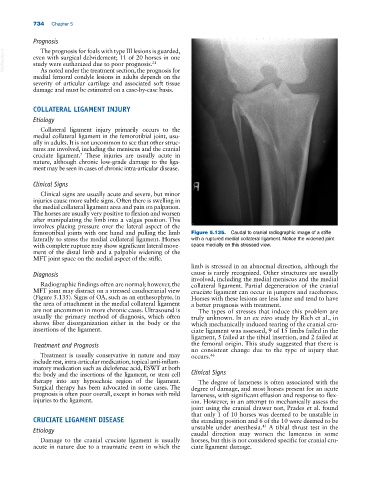
femorotibial joints with one hand and pulling the limb Figure 5.135. Caudal to cranial radiographic image of a stifle
laterally to stress the medial collateral ligament. Horses with a ruptured medial collateral ligament. Notice the widened joint
with complete rupture may show significant lateral move- space medially on this stressed view.
ment of the distal limb and a palpable widening of the
MFT joint space on the medial aspect of the stifle.
limb is stressed in an abnormal direction, although the
Diagnosis cause is rarely recognized. Other structures are usually
involved, including the medial meniscus and the medial
Radiographic findings often are normal; however, the collateral ligament. Partial degeneration of the cranial
MFT joint may distract on a stressed caudocranial view cruciate ligament can occur in jumpers and racehorses.
(Figure 5.135). Signs of OA, such as an enthesophyte, in Horses with these lesions are less lame and tend to have
the area of attachment in the medial collateral ligament a better prognosis with treatment.
are not uncommon in more chronic cases. Ultrasound is The types of stresses that induce this problem are
usually the primary method of diagnosis, which often truly unknown. In an ex vivo study by Rich et al., in
shows fiber disorganization either in the body or the which mechanically induced tearing of the cranial cru-
insertions of the ligament. ciate ligament was assessed, 9 of 15 limbs failed in the
ligament, 5 failed at the tibial insertion, and 2 failed at
Treatment and Prognosis the femoral origin. This study suggested that there is
no consistent change due to the type of injury that
Treatment is usually conservative in nature and may occurs. 46
include rest, intra‐articular medication, topical anti‐inflam-
matory medication such as diclofenac acid, ESWT at both
the body and the insertions of the ligament, or stem cell Clinical Signs
therapy into any hypoechoic region of the ligament. The degree of lameness is often associated with the
Surgical therapy has been advocated in some cases. The degree of damage, and most horses present for an acute
prognosis is often poor overall, except in horses with mild lameness, with significant effusion and response to flex-
injuries to the ligament. ion. However, in an attempt to mechanically assess the
joint using the cranial drawer test, Prades et al. found
that only 1 of 10 horses was deemed to be unstable in
CRUCIATE LIGAMENT DISEASE the standing position and 6 of the 10 were deemed to be
43
Etiology unstable under anesthesia. A tibial thrust test in the
caudal direction may worsen the lameness in some
Damage to the cranial cruciate ligament is usually horses, but this is not considered specific for cranial cru-
acute in nature due to a traumatic event in which the ciate ligament damage.