
Page 764 - Adams and Stashak's Lameness in Horses, 7th Edition
P. 764
730 Chapter 5
VetBooks.ir
A
B C
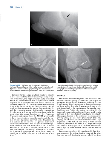
Figure 5.130. (A) Flexed lateral radiograph identifying a fragment was attached to the cranial cruciate ligament, but was
fracture of the caudal aspect of the medial femoral condyle (arrow). loose during arthroscopic examination. (C) A caudal to cranial
(B) A flexed lateromedial radiographic image that demonstrates radiograph demonstrating fragmentation of the intercondylar
fragmentation of the intercondylar eminence of the tibia (arrow). The eminence of the tibia (arrow).
Peroneus tertius origin avulsion fractures usually Treatment
induce flexion of the stifle with corresponding exten-
sion of the hock, and fragments are usually contained Small intra‐articular fragments can be excised and
within the femoropatellar joint. Fragmentation of the removed arthroscopically in most cases. It is important
origin of the long digital extensor muscle can induce to explore the entire joint, both front and back, because
lameness (Figure 5.131), although the author has seen fragments and debris can migrate to the caudal aspect of
these as incidental lesions on routine radiographs of the joint, and to determine if secondary damage to the
yearlings. Fragments can be removed arthroscopically, soft tissues (such as the meniscus and the articular carti-
and the prognosis is good. Radiographs can confirm lage) is present. Proliferative lesions can be found in the
the diagnosis, and ultrasound is sometimes useful in intercondylar area of the tibia and femur, which can be
identifying the exact location of the fragment. debrided if they are clinically significant. Specifically,
Fragments originating from the MICET are thought fibrous proliferation of the soft tissues can be present in
to be avulsion fractures from the insertion of the cra- chronic cases, as can bony proliferation that may
nial cruciate ligament; however, some fragments in impinge on soft tissue structures with movement. Large
48
this location do not involve the ligament insertion. fragments in the MICET that have a stable fibrous
Walmsley demonstrated that the cranial cruciate liga- adhesion often can be left intact. If the fragment is large
ment insertion was not involved in 7 of 12 horses and involves a large portion of the intercondylar emi-
with MICET fractures. Cranial ligament of MM can nence, then lag screw fixation can be used to stabilize
also be damaged. Ultrasound examination is valua- the area. 61
48
ble in assessing prognosis, which can be worsened by Fragment removal should be performed if there is no
significant secondary damage to soft tissues of the compromise to the weight‐bearing aspect of the joint;
joints. 2 however, internal fixation is recommended if the joint